Copyright
©The Author(s) 2023.
World J Clin Cases. May 16, 2023; 11(14): 3195-3203
Published online May 16, 2023. doi: 10.12998/wjcc.v11.i14.3195
Published online May 16, 2023. doi: 10.12998/wjcc.v11.i14.3195
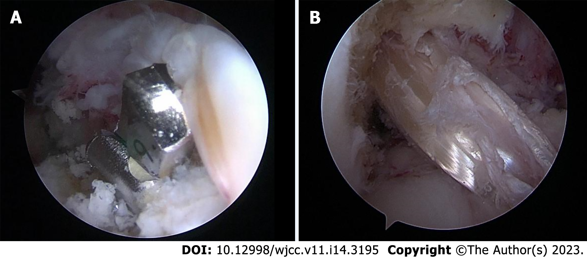
Figure 2 Arthroscopic image.
A: The tibial socket was created at the anatomic tibial site indexing off the anterior horn of the lateral meniscus using a FlipCutter aiming guide (Arthrex); B: Arthroscopic view from the anterolateral portal in the right knee shows that the graft was hoisted up into the socket to the appropriate depth with TightRope shortening strands.
- Citation: An BJ, Wang YT, Zhao Z, Wang MX, Xing GY. Comparative study of the clinical efficacy of all-inside and traditional techniques in anterior cruciate ligament reconstruction. World J Clin Cases 2023; 11(14): 3195-3203
- URL: https://www.wjgnet.com/2307-8960/full/v11/i14/3195.htm
- DOI: https://dx.doi.org/10.12998/wjcc.v11.i14.3195


