Copyright
©The Author(s) 2023.
World J Clin Cases. May 6, 2023; 11(13): 3038-3044
Published online May 6, 2023. doi: 10.12998/wjcc.v11.i13.3038
Published online May 6, 2023. doi: 10.12998/wjcc.v11.i13.3038
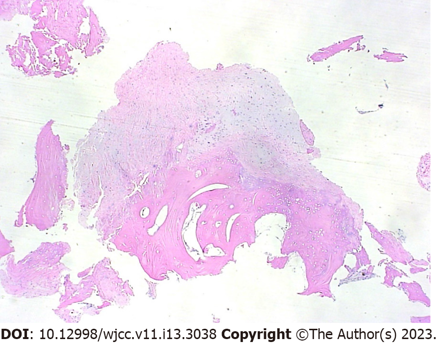
Figure 4 Histopathologic analysis of the hamate bony lesion shows thick cartilaginous tissue, such as the typical cartilaginous cap seen in the common osteochondroma with no malignant changes (hematoxylin-eosin stain, original magnification x 100).
- Citation: Kwon TY, Lee YK. Multiple flexor tendon ruptures due to osteochondroma of the hamate: A case report. World J Clin Cases 2023; 11(13): 3038-3044
- URL: https://www.wjgnet.com/2307-8960/full/v11/i13/3038.htm
- DOI: https://dx.doi.org/10.12998/wjcc.v11.i13.3038


