Copyright
©The Author(s) 2023.
World J Clin Cases. May 6, 2023; 11(13): 3010-3016
Published online May 6, 2023. doi: 10.12998/wjcc.v11.i13.3010
Published online May 6, 2023. doi: 10.12998/wjcc.v11.i13.3010
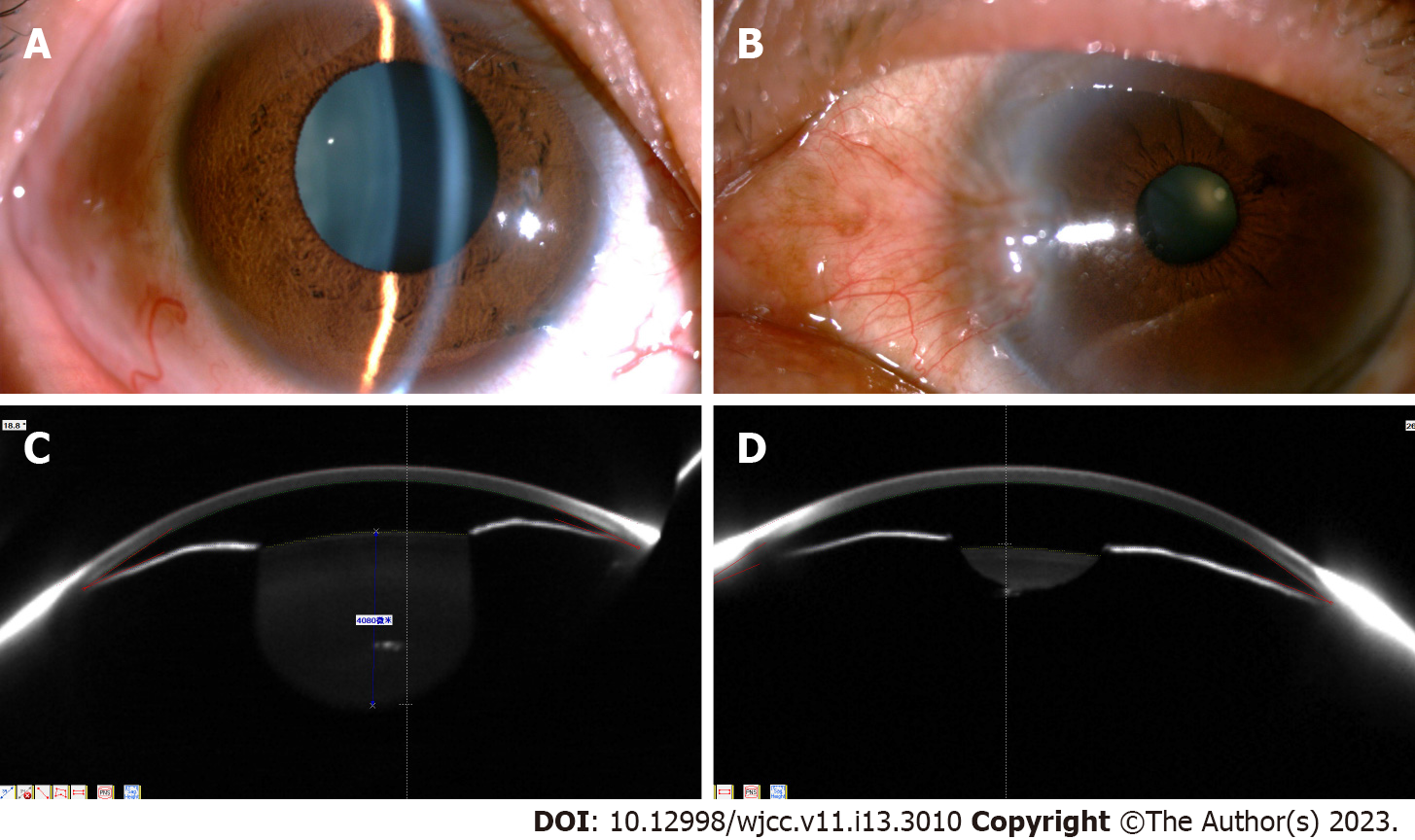
Figure 1 Anterior segment photographs and pentacam analysis.
A and B: Bilateral corneas show transparency. Pupil diameter: OD 6.5 mm and OS 2.8 mm. The hole of iridotomy was marked by a black arrow; B: Different perspectives; C and D: Central and peripheral anterior chambers were shallow, and partial angle closure was present in both eyes.
- Citation: Ma YB, Dang YL. Bilateral malignant glaucoma with bullous keratopathy: A case report. World J Clin Cases 2023; 11(13): 3010-3016
- URL: https://www.wjgnet.com/2307-8960/full/v11/i13/3010.htm
- DOI: https://dx.doi.org/10.12998/wjcc.v11.i13.3010


