Copyright
©The Author(s) 2023.
World J Clin Cases. Apr 26, 2023; 11(12): 2694-2707
Published online Apr 26, 2023. doi: 10.12998/wjcc.v11.i12.2694
Published online Apr 26, 2023. doi: 10.12998/wjcc.v11.i12.2694
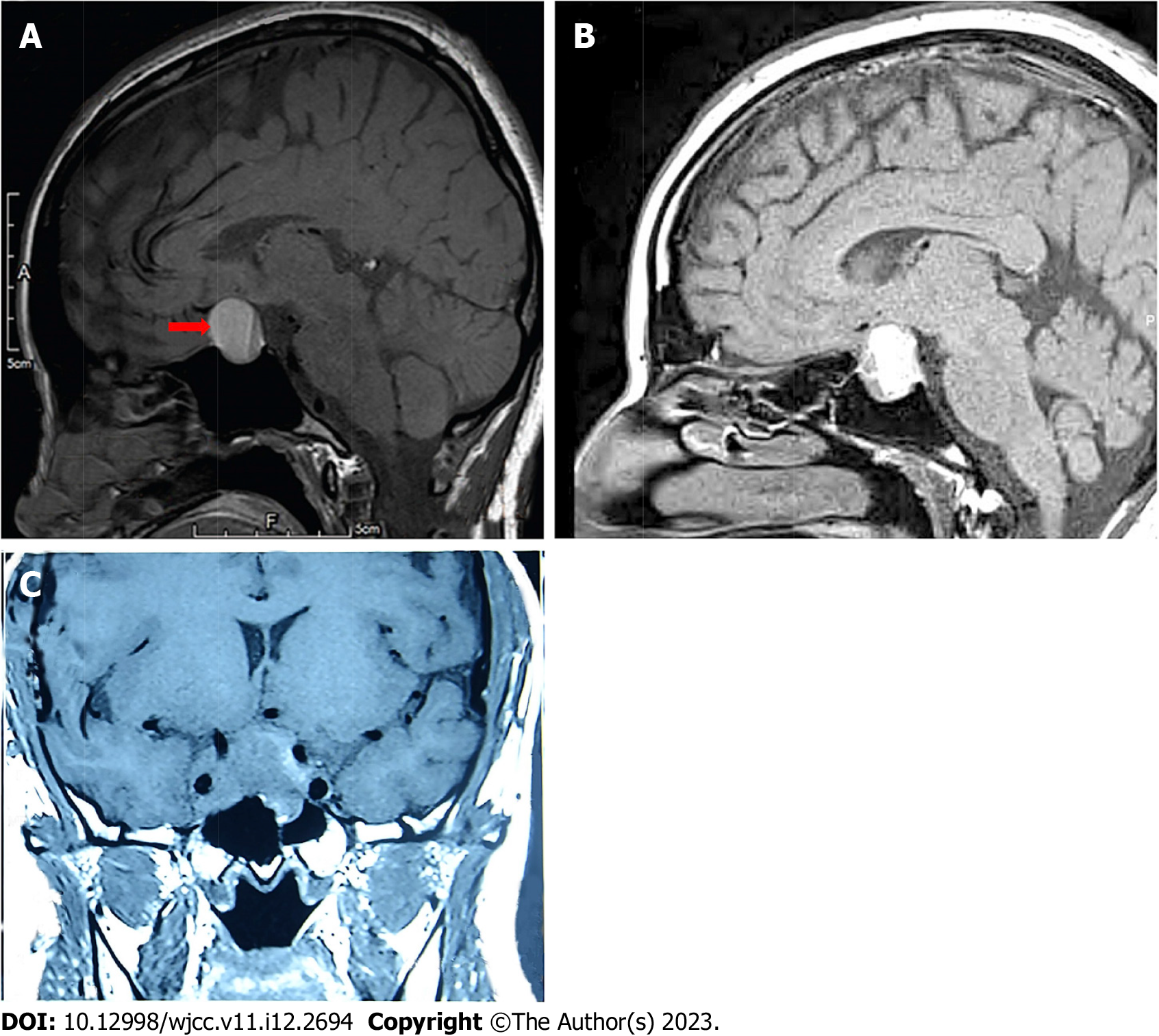
Figure 2 Three typical images of pituitary adenoma apoplexy.
A: Sagittal T1 weighted imaging (T1WI) showed isointensity and hyperintensity with a visible liquid level (Case 15); B: Sagittal T1WI showed mixed intensity (mainly hyperintensity) (Case 40); C: Coronal T1WI showed isointensity (Case 5).
- Citation: Jia XY, Guo XP, Yao Y, Deng K, Lian W, Xing B. Surgical management of pituitary adenoma during pregnancy. World J Clin Cases 2023; 11(12): 2694-2707
- URL: https://www.wjgnet.com/2307-8960/full/v11/i12/2694.htm
- DOI: https://dx.doi.org/10.12998/wjcc.v11.i12.2694


