Copyright
©The Author(s) 2023.
World J Clin Cases. Apr 26, 2023; 11(12): 2637-2656
Published online Apr 26, 2023. doi: 10.12998/wjcc.v11.i12.2637
Published online Apr 26, 2023. doi: 10.12998/wjcc.v11.i12.2637
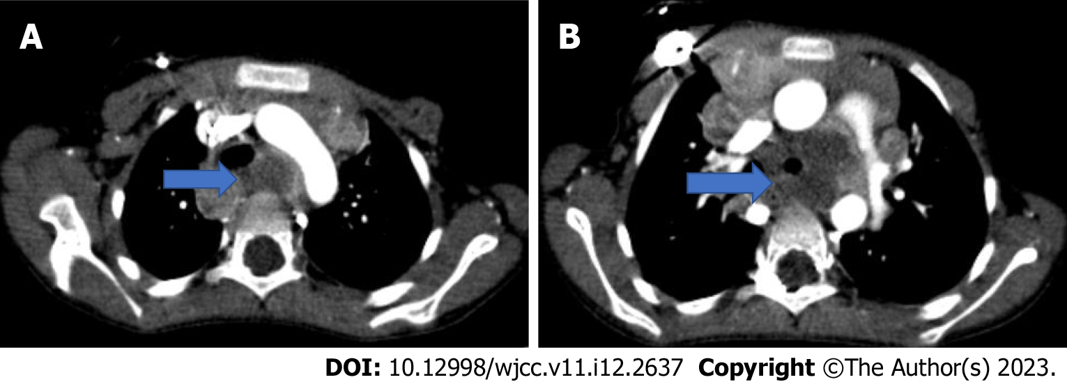
Figure 26 The axial computed tomography images.
A and B: In the axial computed tomography images, necrotic lymphadenopathies (blue arrow) are present in paratracheal, precarinal and subcarinal areas; B: They deviate the trachea anteriorly and invade the left bronchus.
- Citation: Çinar HG, Gulmez AO, Üner Ç, Aydin S. Mediastinal lesions in children. World J Clin Cases 2023; 11(12): 2637-2656
- URL: https://www.wjgnet.com/2307-8960/full/v11/i12/2637.htm
- DOI: https://dx.doi.org/10.12998/wjcc.v11.i12.2637


