Copyright
©The Author(s) 2023.
World J Clin Cases. Apr 26, 2023; 11(12): 2637-2656
Published online Apr 26, 2023. doi: 10.12998/wjcc.v11.i12.2637
Published online Apr 26, 2023. doi: 10.12998/wjcc.v11.i12.2637
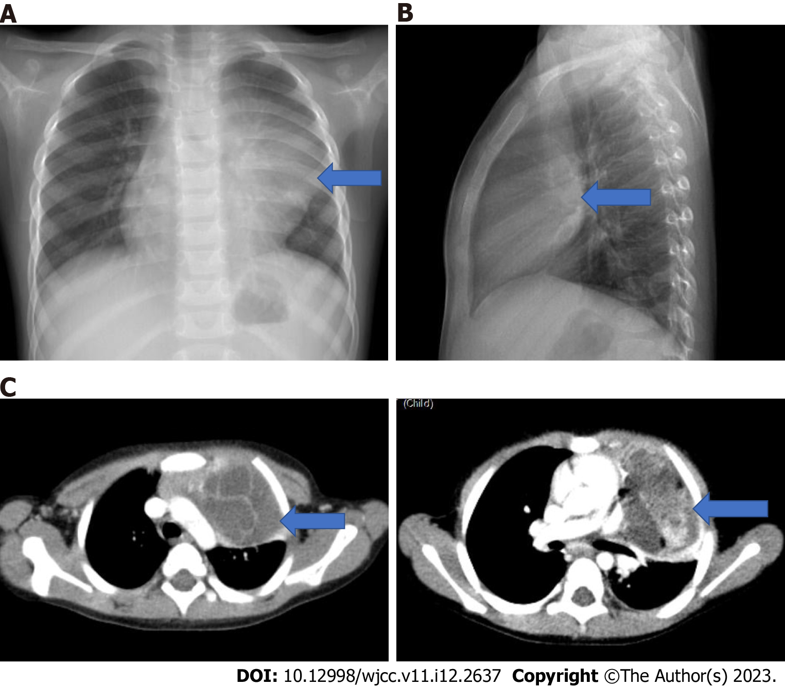
Figure 18 A 3-year-old girl with mature teratoma.
She presents with dyspnea. A: On the posteroanterior chest radiograph the mass (blue arrow) erases the contour of the hilus and heart on the left, slightly compresses the left main bronchus; B: It is located in the anterior mediastinum on the lateral X-ray (blue arrow); C: In the axial computed tomography images of the same patient, a septated mass lesion containing fluid, calcification and fat density (blue arrow) is located in the anterior mediastinum, it compresses the thymus and slightly compresses the left main bronchus. The mass also extends to the left side of the mediastinum.
- Citation: Çinar HG, Gulmez AO, Üner Ç, Aydin S. Mediastinal lesions in children. World J Clin Cases 2023; 11(12): 2637-2656
- URL: https://www.wjgnet.com/2307-8960/full/v11/i12/2637.htm
- DOI: https://dx.doi.org/10.12998/wjcc.v11.i12.2637


