
















©The Author(s) 2023.
World J Clin Cases. Apr 26, 2023; 11(12): 2637-2656
Published online Apr 26, 2023. doi: 10.12998/wjcc.v11.i12.2637
Published online Apr 26, 2023. doi: 10.12998/wjcc.v11.i12.2637
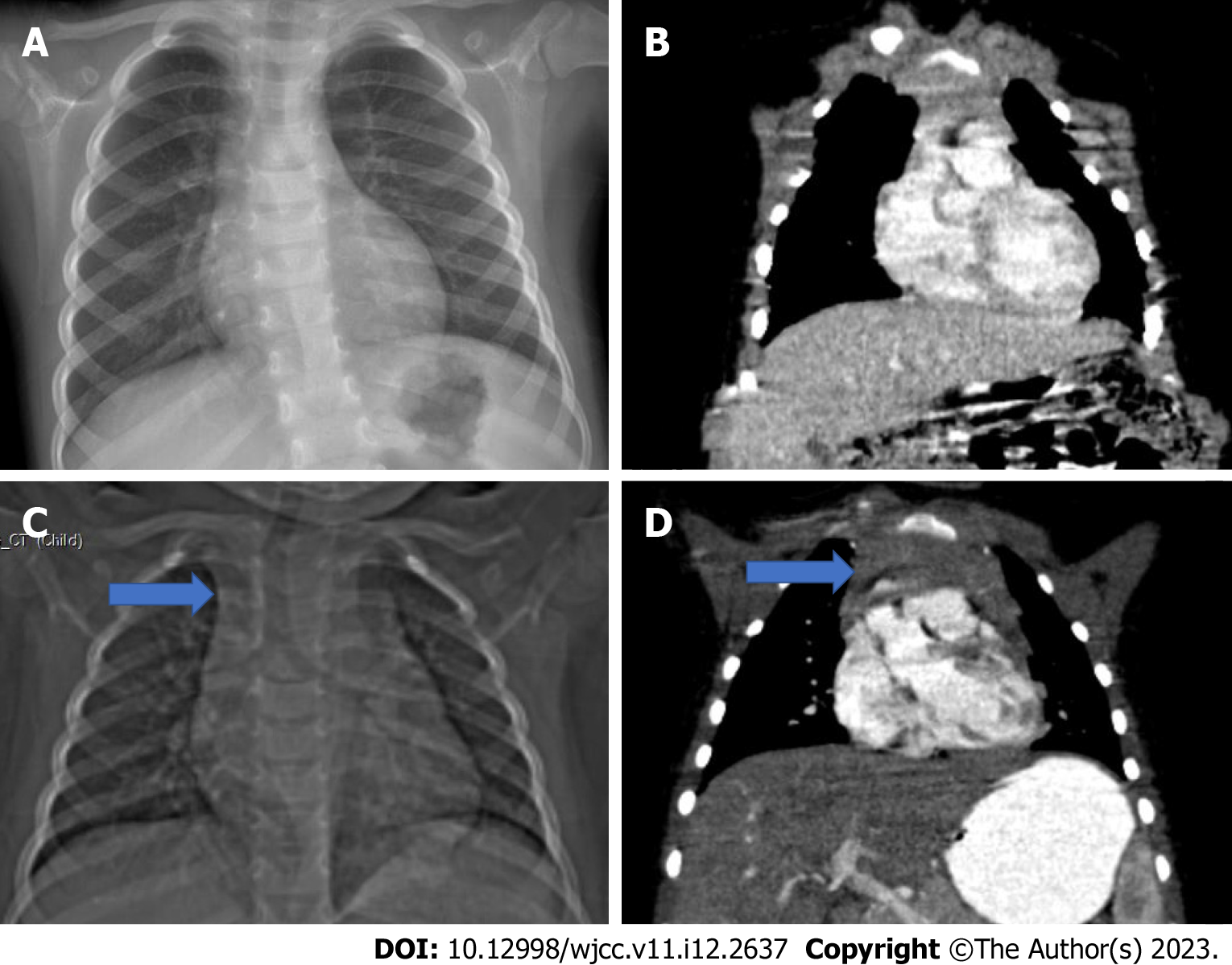
Figure 11 A 3-year-old patient who was followed up with an operated Wilms tumor during chemotherapy.
A: The thymus tissue is observed to be smaller than normal in the posteroanterior chest radiograph; B: Coronal computed tomography examination; C and D: In the second month follow-up of the same patient after the end of chemotherapy. Hyperplasia in the thymus (blue arrow) is observed in the scout (C) and coronal plane computed tomography images (D).
- Citation: Çinar HG, Gulmez AO, Üner Ç, Aydin S. Mediastinal lesions in children. World J Clin Cases 2023; 11(12): 2637-2656
- URL: https://www.wjgnet.com/2307-8960/full/v11/i12/2637.htm
- DOI: https://dx.doi.org/10.12998/wjcc.v11.i12.2637