Copyright
©The Author(s) 2023.
World J Clin Cases. Apr 26, 2023; 11(12): 2637-2656
Published online Apr 26, 2023. doi: 10.12998/wjcc.v11.i12.2637
Published online Apr 26, 2023. doi: 10.12998/wjcc.v11.i12.2637
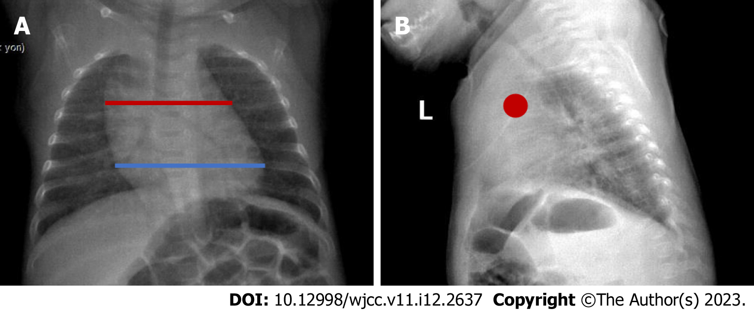
Figure 3 A 3-year-old boy normal thymus.
A: The red line shows the thymus and the blue line shows the heart in the posteroanterior chest radiograph. In infants, the thymus erases the heart contours, and because it is superposed with the hiluses, the hiluses cannot be clearly distinguished. It also does not create a compression effect. B: Anterior location of the thymus is observed with a red dot on the lateral radiograph.
- Citation: Çinar HG, Gulmez AO, Üner Ç, Aydin S. Mediastinal lesions in children. World J Clin Cases 2023; 11(12): 2637-2656
- URL: https://www.wjgnet.com/2307-8960/full/v11/i12/2637.htm
- DOI: https://dx.doi.org/10.12998/wjcc.v11.i12.2637


