
















©The Author(s) 2023.
World J Clin Cases. Apr 16, 2023; 11(11): 2567-2575
Published online Apr 16, 2023. doi: 10.12998/wjcc.v11.i11.2567
Published online Apr 16, 2023. doi: 10.12998/wjcc.v11.i11.2567
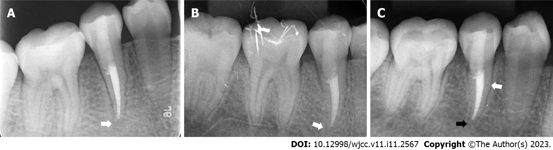
Figure 5 Follow-up of the right mandibular second premolar.
A: 3-mo follow-up. The hypodense area around the root apex was significantly diminished (white arrow); B: 6-mo follow-up. The periapical hypodense area was completely healed (white arrow); C: 12-mo follow-up. The periodontal ligament was narrowed (white arrow). Complete closure of the apex was observed, and there was an obvious calcification layer generated just around the apical barrier materials (black arrow).
- Citation: Chai R, Yang X, Zhang AS. Different endodontic treatments induced root development of two nonvital immature teeth in the same patient: A case report. World J Clin Cases 2023; 11(11): 2567-2575
- URL: https://www.wjgnet.com/2307-8960/full/v11/i11/2567.htm
- DOI: https://dx.doi.org/10.12998/wjcc.v11.i11.2567