Copyright
©The Author(s) 2023.
World J Clin Cases. Apr 6, 2023; 11(10): 2308-2314
Published online Apr 6, 2023. doi: 10.12998/wjcc.v11.i10.2308
Published online Apr 6, 2023. doi: 10.12998/wjcc.v11.i10.2308
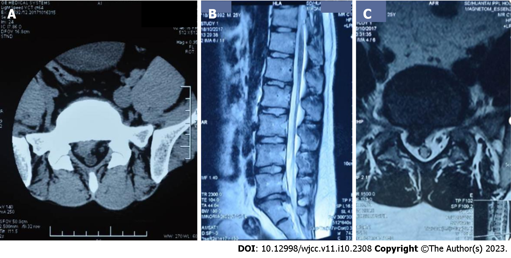
Figure 1 Computed tomography scan of lumbar prolapsed intraforaminal right-sided L5/S1 disc obtained in 2017.
A: Transverse position: L5/S1 disc herniation; B: Sagittal position: L5/S1 disc herniation; C: Nucleus pulposus detached into spinal canal, right nerve root compression.
- Citation: Wang CA, Zhao HF, Ju J, Kong L, Sun CJ, Zheng YK, Zhang F, Hou GJ, Guo CC, Cao SN, Wang DD, Shi B. Reabsorption of intervertebral disc prolapse after conservative treatment with traditional Chinese medicine: A case report. World J Clin Cases 2023; 11(10): 2308-2314
- URL: https://www.wjgnet.com/2307-8960/full/v11/i10/2308.htm
- DOI: https://dx.doi.org/10.12998/wjcc.v11.i10.2308


