















©The Author(s) 2023.
World J Clin Cases. Apr 6, 2023; 11(10): 2260-2266
Published online Apr 6, 2023. doi: 10.12998/wjcc.v11.i10.2260
Published online Apr 6, 2023. doi: 10.12998/wjcc.v11.i10.2260
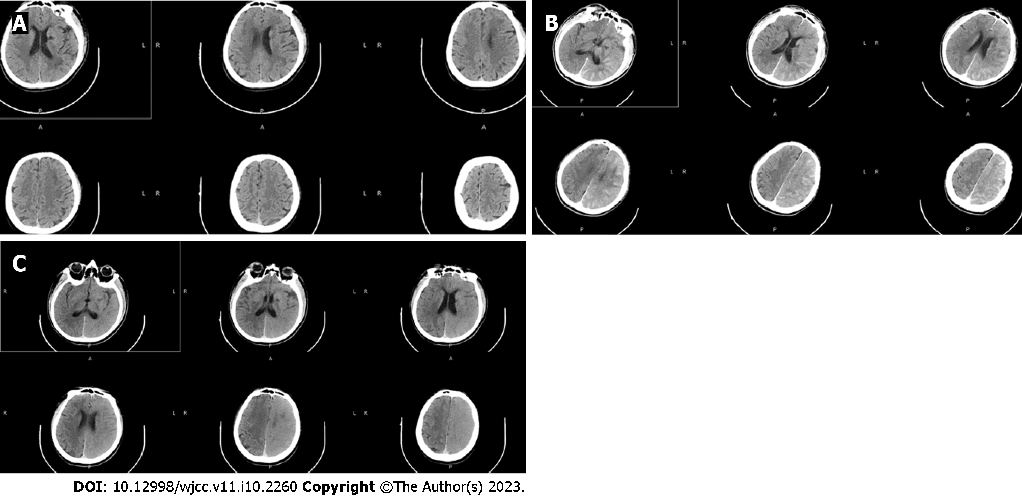
Figure 2 Computed tomography of the brain before and 2 h and 7 h after the procedure.
A: The normal occipital lobe of the left cerebral hemisphere on the preprocedure computed Tomography (CT); B: The increased density of the interhemispheric cistern, left fissure cistern and part of the left sulcus on CT 2 h after digital subtraction angiography (DSA); C: Seven hours after DSA, indicating the swelling of the left frontal, temporal, parietal and occipital lobe brain tissue, the disappearance of the high-density shadow of sulci and gyri, the shallow appearance of the sulci and gyri, and the higher density of the left cerebral hemisphere than the right cerebral hemisphere.
- Citation: Zhang ZY, Lv H, Wang PJ, Zhao DY, Zhang LY, Wang JY, Hao JH. Unilateral contrast-induced encephalopathy with contrast medium exudation: A case report. World J Clin Cases 2023; 11(10): 2260-2266
- URL: https://www.wjgnet.com/2307-8960/full/v11/i10/2260.htm
- DOI: https://dx.doi.org/10.12998/wjcc.v11.i10.2260