
















©The Author(s) 2023.
World J Clin Cases. Jan 6, 2023; 11(1): 30-46
Published online Jan 6, 2023. doi: 10.12998/wjcc.v11.i1.30
Published online Jan 6, 2023. doi: 10.12998/wjcc.v11.i1.30
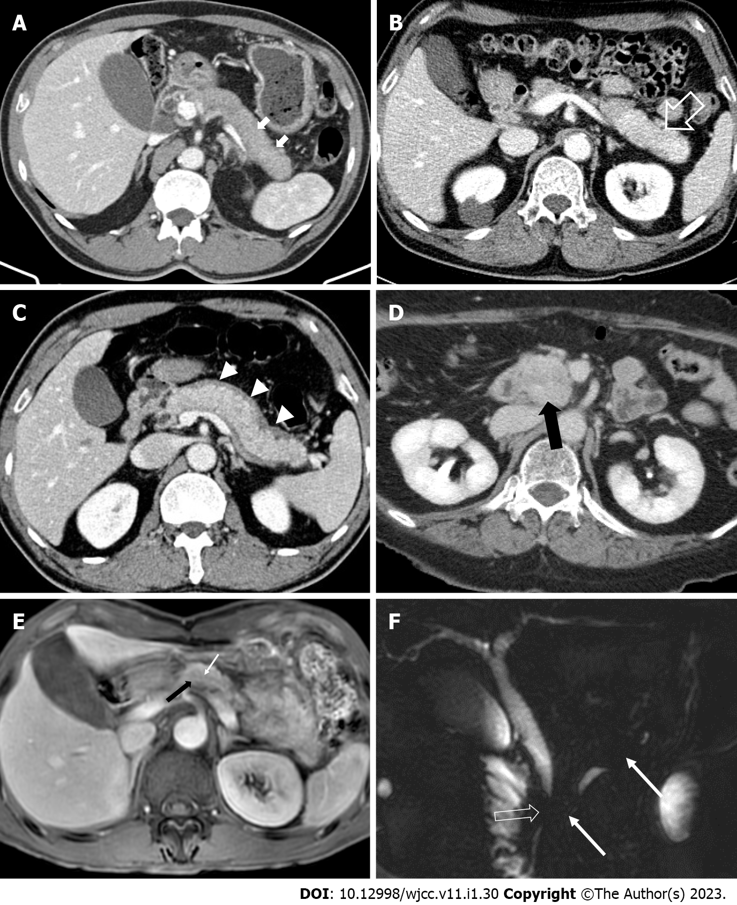
Figure 2 Computed tomography and magnetic resonance imaging findings of autoimmune pancreatitis.
A: Axial computed tomography (CT) image shows “sausage-like appearance” of diffuse enlargement of the pancreas with absence of pancreatic clefts (white arrows); B: Delayed enhancement (open white arrow) with swelling of the pancreatic tail; C: Hypotonic band (white arrow heads), called the “capsule-like rim”, around the pancreas; D: Axial CT image demonstrates a homogenous enhanced mass (black arrow) with proven autoimmune pancreatitis in the pancreas head mimicking pancreatic ductal adenocarcinoma; E: A pancreatic-phase dynamic contrast-enhanced magnetic resonance imaging (MRI) shows a mild arterial enhanced mass-like lesion (black arrow) with a penetrating duct sign (white arrow); F: An MRI cholangiopancreatography image shows irregular narrowing of the main pancreatic duct (white arrows) without upstream dilatation and short segment stricture of the common bile duct (open white arrow).
- Citation: Kim SH, Lee YC, Chon HK. Challenges for clinicians treating autoimmune pancreatitis: Current perspectives. World J Clin Cases 2023; 11(1): 30-46
- URL: https://www.wjgnet.com/2307-8960/full/v11/i1/30.htm
- DOI: https://dx.doi.org/10.12998/wjcc.v11.i1.30