Copyright
©The Author(s) 2023.
World J Clin Cases. Jan 6, 2023; 11(1): 135-142
Published online Jan 6, 2023. doi: 10.12998/wjcc.v11.i1.135
Published online Jan 6, 2023. doi: 10.12998/wjcc.v11.i1.135
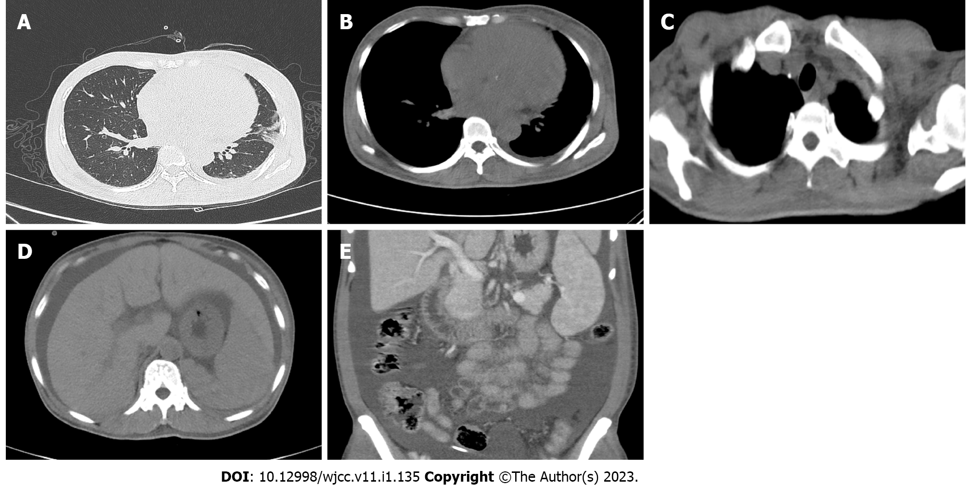
Figure 1 Images from chest and abdominal computed tomography performed on the patient for the first time on March 16, 2021.
A: Inflammatory lesions in the lingual segment of the left lung and lower lobes of both lungs and slightly thickened bilateral pleura and a small amount of effusion in the left pleural cavity; B: Slight enlargement of the heart and enlarged lymph nodes in the mediastinum; C: Enlarged lymph nodes in the bilateral axilla; D: Widened liver fissure and abdominal hydrops; E: Splenomegaly, pelvic hydrops, swollen and thick intestinal walls, and blurred fat space in the abdominal and pelvic cavity.
- Citation: Zhou XL, Chang YH, Li L, Ren J, Wu XL, Zhang X, Wu P, Tang SH. Polyneuropathy organomegaly endocrinopathy M-protein and skin changes syndrome with ascites as an early-stage manifestation: A case report. World J Clin Cases 2023; 11(1): 135-142
- URL: https://www.wjgnet.com/2307-8960/full/v11/i1/135.htm
- DOI: https://dx.doi.org/10.12998/wjcc.v11.i1.135


