
















©The Author(s) 2023.
World J Clin Cases. Jan 6, 2023; 11(1): 116-126
Published online Jan 6, 2023. doi: 10.12998/wjcc.v11.i1.116
Published online Jan 6, 2023. doi: 10.12998/wjcc.v11.i1.116
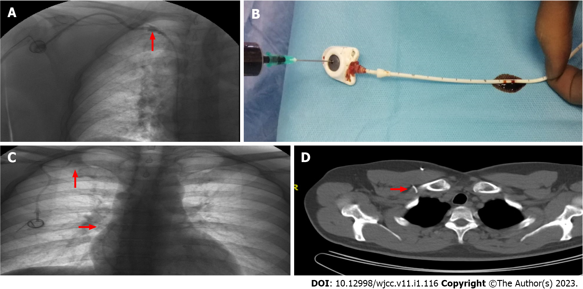
Figure 1 Rupture of a catheter implanted through the right subclavian vein.
A: Fluoroscopy revealed contrast extravasation (red arrow) in patient 1; B: In patient 1, at the ex situ evaluation of the catheter, we found an approximately 2 cm linear breakage. This breakage was 9 cm distal to the port-catheter attachment point; C: In patient 2, radiological evaluation revealed a complete breakage, and the distal end of the catheter was found to be free floating in the atrium (red arrow); D: The port site was between the clavicle and the first rib (red arrow) in patient 2. The interventional radiology team removed the broken distal end through an angiographic intervention.
- Citation: Erdemir A, Rasa HK. Impact of central venous port implantation method and access choice on outcomes. World J Clin Cases 2023; 11(1): 116-126
- URL: https://www.wjgnet.com/2307-8960/full/v11/i1/116.htm
- DOI: https://dx.doi.org/10.12998/wjcc.v11.i1.116