Copyright
©The Author(s) 2022.
World J Clin Cases. Mar 26, 2022; 10(9): 2938-2947
Published online Mar 26, 2022. doi: 10.12998/wjcc.v10.i9.2938
Published online Mar 26, 2022. doi: 10.12998/wjcc.v10.i9.2938
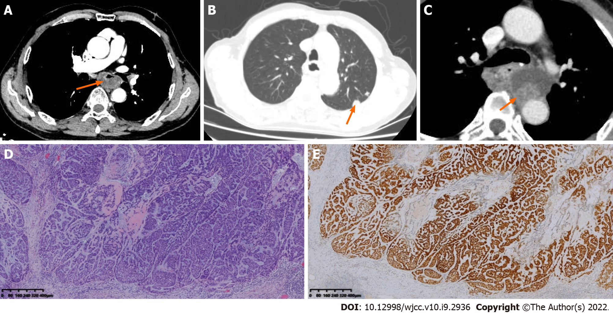
Figure 3 Computed tomography images, hematoxylin and eosin staining, and SOX-10 immunohistochemistry of patient 3.
A: Enhanced computed tomography (CT) showed thickening with eccentric stenosis of the middle esophagus, with a complete mucosal layer and cystic change or necrosis(orange arrow); B: At the four-month review, chest CT revealed multiple lung metastases (orange arrow); C: At the four-month review, enhanced CT revealed a cystic-solid mass (orange arrow) near the anastomosis; D: HE staining showed mainly epithelioid cells with hyperchromatic and pleomorphic nuclei and infiltrative growth toward the periphery. (Magnification × 40); E: Immunohistochemistry showing the expression of SOX-10. (Magnification × 40).
- Citation: Lu H, Zhao HP, Liu YY, Yu J, Wang R, Gao JB. Esophageal myoepithelial carcinoma: Four case reports. World J Clin Cases 2022; 10(9): 2938-2947
- URL: https://www.wjgnet.com/2307-8960/full/v10/i9/2938.htm
- DOI: https://dx.doi.org/10.12998/wjcc.v10.i9.2938


