Copyright
©The Author(s) 2022.
World J Clin Cases. Mar 26, 2022; 10(9): 2938-2947
Published online Mar 26, 2022. doi: 10.12998/wjcc.v10.i9.2938
Published online Mar 26, 2022. doi: 10.12998/wjcc.v10.i9.2938
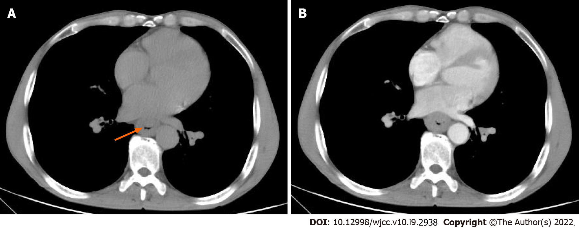
Figure 1 Chest computed tomography images of patient 1.
A: Unenhanced computed tomography shows local thickening and luminal narrowing of the esophagus (orange arrow) with an evident fat space between the lesion and surrounding tissues; B: After contrast injection, the mass showed mild homogeneous enhancement with no cystic changes or necrosis.
- Citation: Lu H, Zhao HP, Liu YY, Yu J, Wang R, Gao JB. Esophageal myoepithelial carcinoma: Four case reports. World J Clin Cases 2022; 10(9): 2938-2947
- URL: https://www.wjgnet.com/2307-8960/full/v10/i9/2938.htm
- DOI: https://dx.doi.org/10.12998/wjcc.v10.i9.2938


