
















©The Author(s) 2022.
World J Clin Cases. Mar 26, 2022; 10(9): 2871-2877
Published online Mar 26, 2022. doi: 10.12998/wjcc.v10.i9.2871
Published online Mar 26, 2022. doi: 10.12998/wjcc.v10.i9.2871
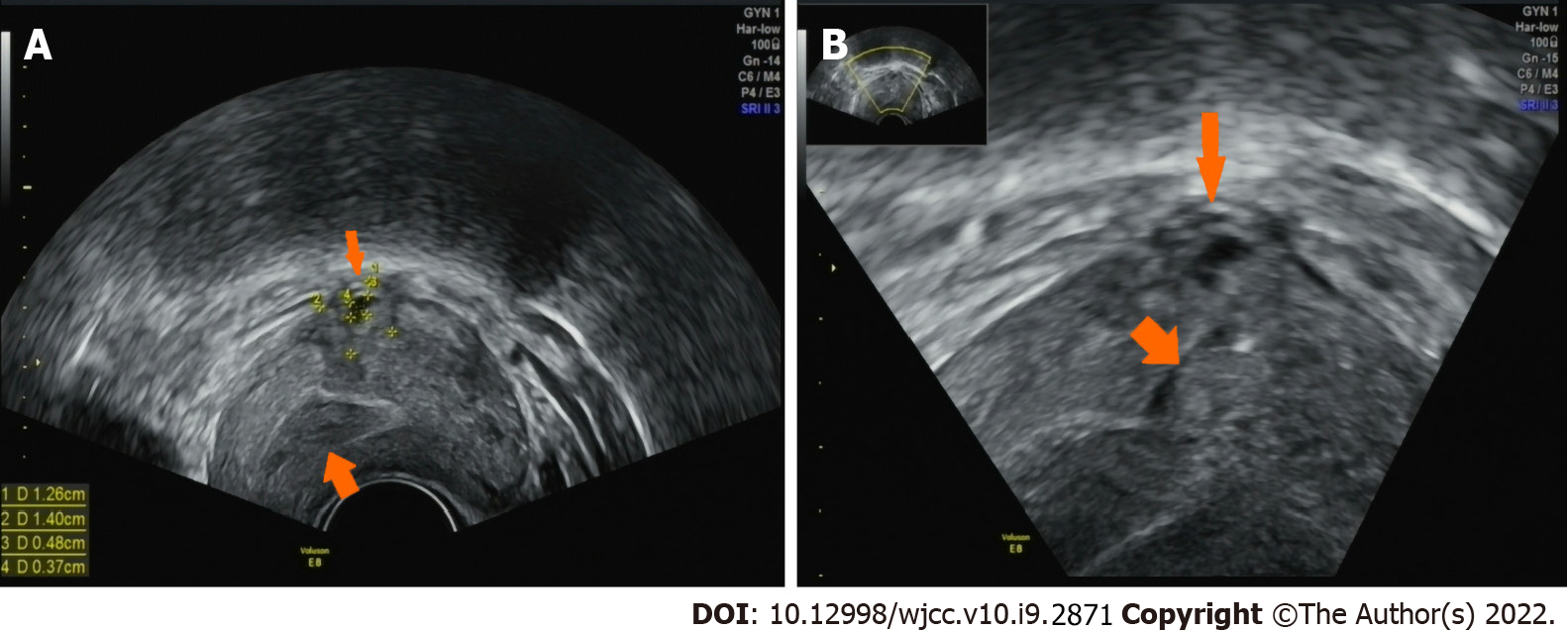
Figure 1 Transvaginal ultrasound image of intrauterine pregnancy at day 33 after embryo transfer.
A: The transvaginal ultrasound showed a heterogeneous echogenic mass measuring 1.40 cm × 1.26 cm in size slightly arising from the left corner of the uterus, which had a 0.48 cm × 0.37 cm anechoic region inside (thin arrow). The endometrial–myometrial junction was displayed (coarse arrow); B: The transvaginal ultrasound showed a slender and extremely hypoechoic area stretching to the uterine cavity (coarse arrow) and the serosal surface of the heterogeneous echogenic mass was covered by feeble myometrial tissue (thin arrow).
- Citation: Xie QJ, Li X, Ni DY, Ji H, Zhao C, Ling XF. Intramural pregnancy after in vitro fertilization and embryo transfer: A case report. World J Clin Cases 2022; 10(9): 2871-2877
- URL: https://www.wjgnet.com/2307-8960/full/v10/i9/2871.htm
- DOI: https://dx.doi.org/10.12998/wjcc.v10.i9.2871