Copyright
©The Author(s) 2022.
World J Clin Cases. Mar 6, 2022; 10(7): 2294-2300
Published online Mar 6, 2022. doi: 10.12998/wjcc.v10.i7.2294
Published online Mar 6, 2022. doi: 10.12998/wjcc.v10.i7.2294
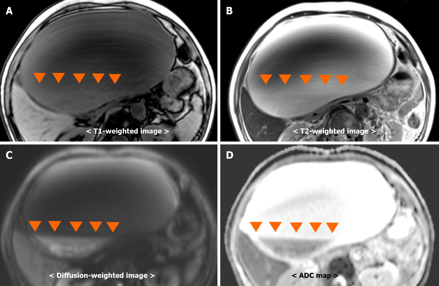
Figure 3 Simple abdominal magnetic resonance imaging.
Fluid-fluid levels (orange arrowheads) from debris in the hepatic cyst. Debris observed as, A: Hyperintensity on a T1-weighted image; B: Hyperintensity on a T2-weighted image; C: Hyperintensity on a diffusion-weighted image; D: Hypointensity on an ADC map.
- Citation: Kenzaka T, Sato Y, Nishisaki H. Giant infected hepatic cyst causing exclusion pancreatitis: A case report. World J Clin Cases 2022; 10(7): 2294-2300
- URL: https://www.wjgnet.com/2307-8960/full/v10/i7/2294.htm
- DOI: https://dx.doi.org/10.12998/wjcc.v10.i7.2294


