Copyright
©The Author(s) 2022.
World J Clin Cases. Mar 6, 2022; 10(7): 2294-2300
Published online Mar 6, 2022. doi: 10.12998/wjcc.v10.i7.2294
Published online Mar 6, 2022. doi: 10.12998/wjcc.v10.i7.2294
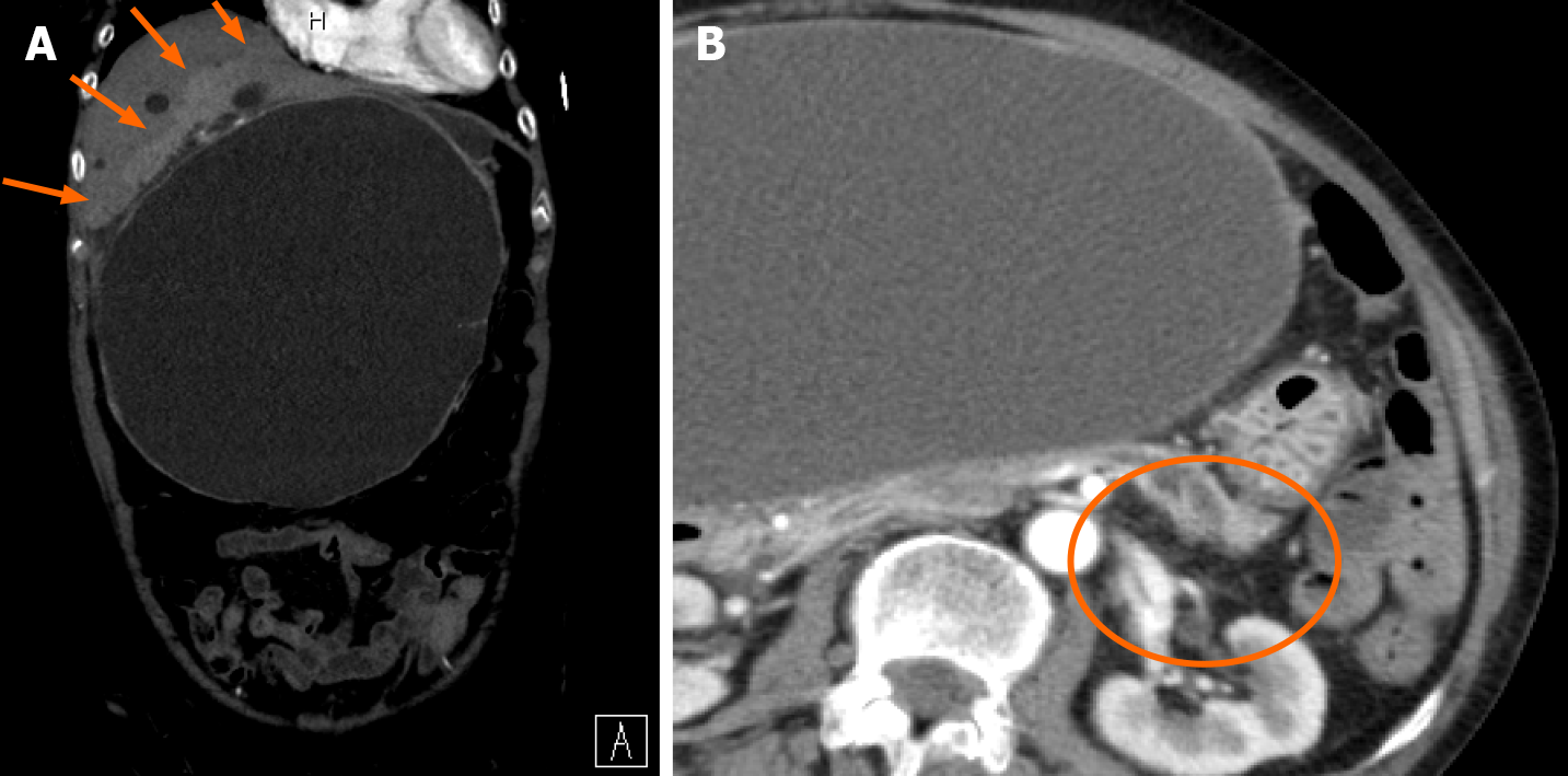
Figure 2 Abdominal dynamic computed tomography (arterial phase) on hospital day 1.
A: A contrast effect is observed in the liver parenchyma around the hepatic cyst (orange arrows). There is no ring enhancement indicating an abscess. The maximum cyst diameter is 203 mm; B: Slight dilation of the main pancreatic duct at the pancreatic tail (orange circle).
- Citation: Kenzaka T, Sato Y, Nishisaki H. Giant infected hepatic cyst causing exclusion pancreatitis: A case report. World J Clin Cases 2022; 10(7): 2294-2300
- URL: https://www.wjgnet.com/2307-8960/full/v10/i7/2294.htm
- DOI: https://dx.doi.org/10.12998/wjcc.v10.i7.2294


