Copyright
©The Author(s) 2022.
World J Clin Cases. Mar 6, 2022; 10(7): 2206-2215
Published online Mar 6, 2022. doi: 10.12998/wjcc.v10.i7.2206
Published online Mar 6, 2022. doi: 10.12998/wjcc.v10.i7.2206
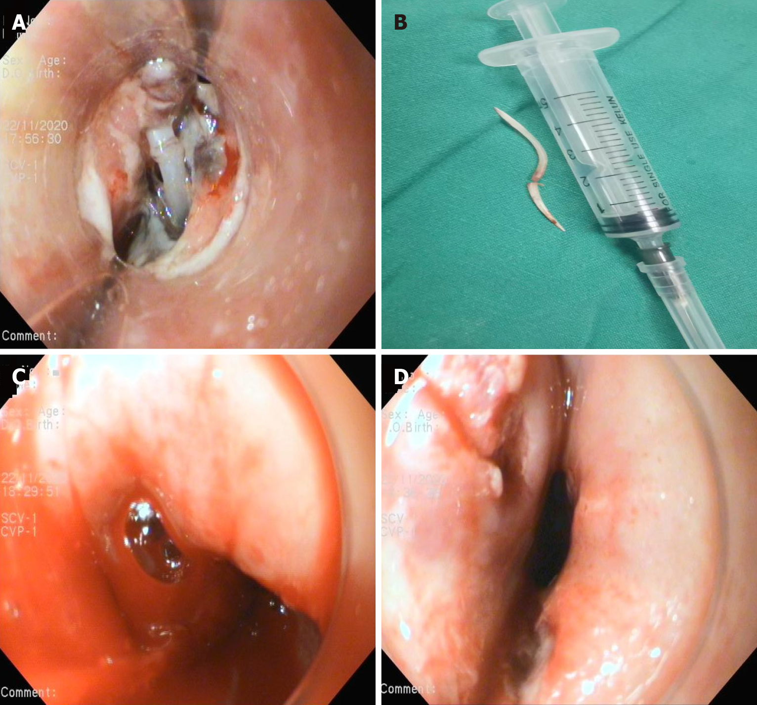
Figure 5 Esophageal view during Esophagogastroduodenoscopy.
A: Both ends of the fishbone inserted into the esophageal wall, 28 cm from the incisors; B: The endoscopically removed fishbone; C: Active blood spurting was noted in the esophageal defect after removal of the fishbone; D: Two longitudinal ulcers were present.
- Citation: Gong H, Wei W, Huang Z, Hu Y, Liu XL, Hu Z. Endovascular stent-graft treatment for aortoesophageal fistula induced by an esophageal fishbone: Two cases report. World J Clin Cases 2022; 10(7): 2206-2215
- URL: https://www.wjgnet.com/2307-8960/full/v10/i7/2206.htm
- DOI: https://dx.doi.org/10.12998/wjcc.v10.i7.2206


