Copyright
©The Author(s) 2022.
World J Clin Cases. Mar 6, 2022; 10(7): 2206-2215
Published online Mar 6, 2022. doi: 10.12998/wjcc.v10.i7.2206
Published online Mar 6, 2022. doi: 10.12998/wjcc.v10.i7.2206
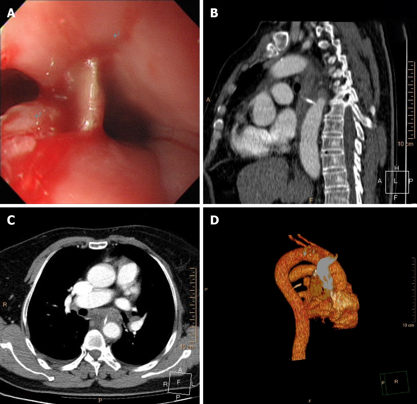
Figure 1 Esophagogastroduodenoscopy and chest computed tomography angiography.
A: Esophagogastroduodenoscopy shows a straight fishbone penetrating the wall of the esophagus, with overflowing purulent secretion, 25 cm from the incisors. The other three pictures reveal preoperative chest computed tomography angiography of the fishbone (length 2.0 cm) penetrating the wall of the esophagus and into the thoracic aorta; B: Sagittal view; C: Axial view; D: Three-dimensional reconstruction.
- Citation: Gong H, Wei W, Huang Z, Hu Y, Liu XL, Hu Z. Endovascular stent-graft treatment for aortoesophageal fistula induced by an esophageal fishbone: Two cases report. World J Clin Cases 2022; 10(7): 2206-2215
- URL: https://www.wjgnet.com/2307-8960/full/v10/i7/2206.htm
- DOI: https://dx.doi.org/10.12998/wjcc.v10.i7.2206


