
















©The Author(s) 2022.
World J Clin Cases. Feb 26, 2022; 10(6): 2023-2029
Published online Feb 26, 2022. doi: 10.12998/wjcc.v10.i6.2023
Published online Feb 26, 2022. doi: 10.12998/wjcc.v10.i6.2023
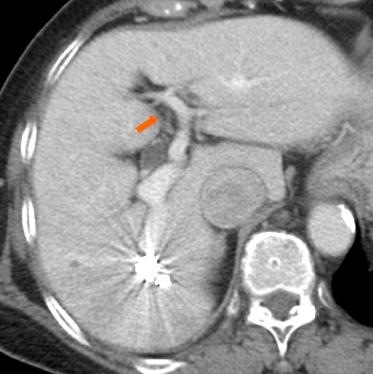
Figure 3 An axial contrast-enhanced computed tomography image that was obtained one week after the procedure reveals sufficient embolization of the intrahepatic portosystemic shunt and expansion of the left intrahepatic portal vein (arrow).
- Citation: Saito H, Murata S, Sugihara F, Ueda T, Yasui D, Miki I, Hayashi H, Kumita SI. Successful embolization of an intrahepatic portosystemic shunt using balloon-occluded retrograde transvenous obliteration: A case report. World J Clin Cases 2022; 10(6): 2023-2029
- URL: https://www.wjgnet.com/2307-8960/full/v10/i6/2023.htm
- DOI: https://dx.doi.org/10.12998/wjcc.v10.i6.2023