Copyright
©The Author(s) 2022.
World J Clin Cases. Feb 26, 2022; 10(6): 1998-2006
Published online Feb 26, 2022. doi: 10.12998/wjcc.v10.i6.1998
Published online Feb 26, 2022. doi: 10.12998/wjcc.v10.i6.1998
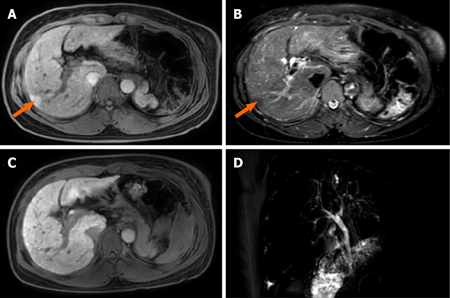
Figure 1 Liver magnetic resonance images.
A: T1-weighted image. The size and shape of the liver are normal, and patchy low signal intensity can be seen in the SVII segment, with a diameter of approximately 15 mm. Loss of spleen; B: T2W spectral attenuated inversion recovery image. The interstitium increased in the liver, and the lesions in the SVII segment showed high signal intensity; C: Gadolinium-ethoxybenzyl-diethylenetriamine penta-acetic acid 15MIN_Delay image. The signal intensity of SVII segment lesions was basically consistent with that of normal liver; D: Magnetic resonance cholangiopancreatography image. No stricture or dilatation of the intrahepatic or extrahepatic bile duct was observed.
- Citation: Liu TF, He JJ, Wang L, Zhang LY. Novel ABCB4 mutations in an infertile female with progressive familial intrahepatic cholestasis type 3: A case report. World J Clin Cases 2022; 10(6): 1998-2006
- URL: https://www.wjgnet.com/2307-8960/full/v10/i6/1998.htm
- DOI: https://dx.doi.org/10.12998/wjcc.v10.i6.1998


