Copyright
©The Author(s) 2022.
World J Clin Cases. Feb 26, 2022; 10(6): 1946-1951
Published online Feb 26, 2022. doi: 10.12998/wjcc.v10.i6.1946
Published online Feb 26, 2022. doi: 10.12998/wjcc.v10.i6.1946
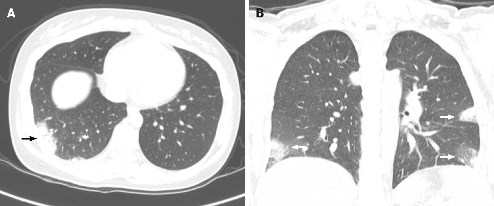
Figure 2 Computed tomography findings.
A: Focal patchy consolidation and peripheral ground-glass opacities (GGOs) of posterior and lateral basal segments of the right lower lobe at first onset (black arrow); B: Interval developed multifocal patchy consolidation and GGOs at anterior and lateral basal segments of the right lower lobe, lateral basal segment of the left lower lobe, and lingular segment of the left upper lobe at second onset of coronal section (white arrow).
- Citation: Lee YJ, Kim YS. Cryptogenic organizing pneumonia associated with pregnancy: A case report. World J Clin Cases 2022; 10(6): 1946-1951
- URL: https://www.wjgnet.com/2307-8960/full/v10/i6/1946.htm
- DOI: https://dx.doi.org/10.12998/wjcc.v10.i6.1946


