Copyright
©The Author(s) 2022.
World J Clin Cases. Feb 26, 2022; 10(6): 1937-1945
Published online Feb 26, 2022. doi: 10.12998/wjcc.v10.i6.1937
Published online Feb 26, 2022. doi: 10.12998/wjcc.v10.i6.1937
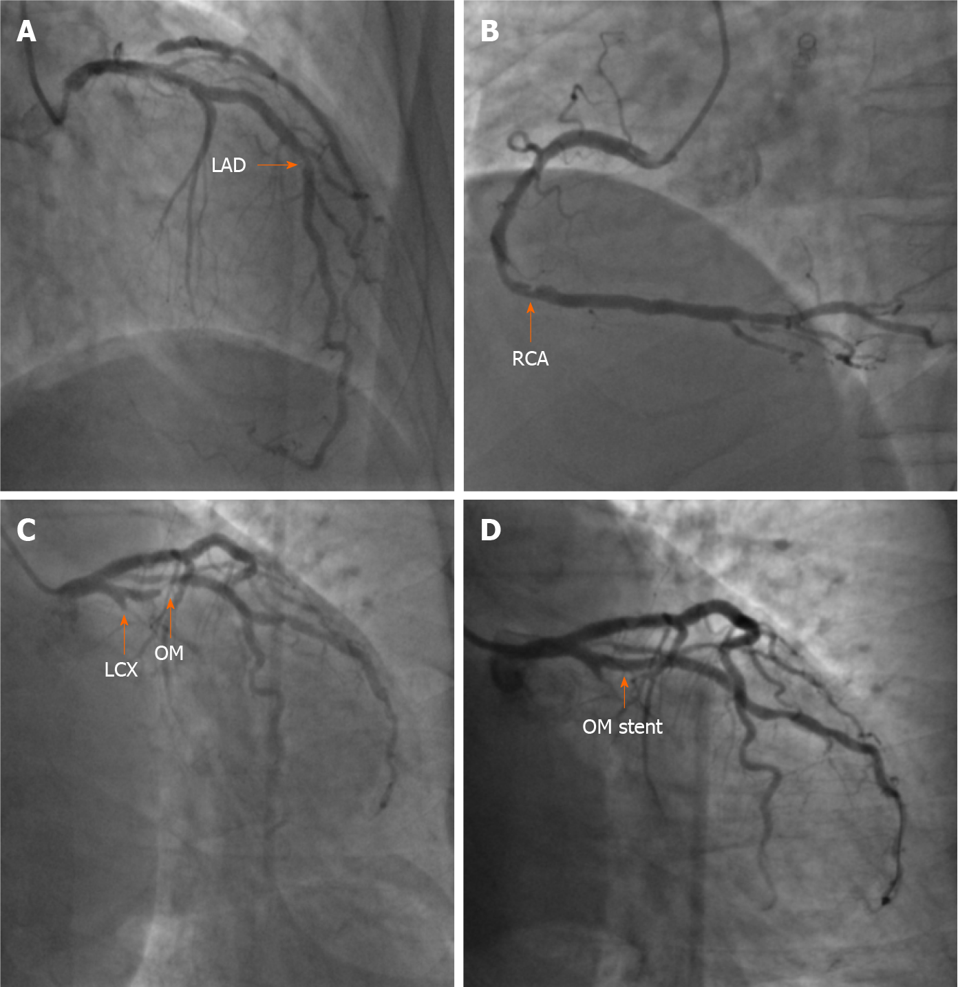
Figure 2 Coronary angiogram and stent implantation.
A: Coronary angiogram showing 60%-70% in-stent restenosis at the middle left anterior descending artery (orange arrow); B: Coronary angiogram showing 60%-70% stenosis of the distal right coronary artery (orange arrow); C: Coronary angiogram showing total occlusion of the proximal left circumflex artery and subtotal occlusion at the opening of the first obtuse marginal branch (OM) (orange arrow); D: After deployment of a stent in the OM, stenosis was eliminated (orange arrow). LAD: Left anterior descending artery; LCX: Left circumflex artery; OM: Obtuse marginal branch; RCA: Right coronary artery.
- Citation: Shi F, Zhang Y, Sun LX, Long S. Life-threatening subclavian artery bleeding following percutaneous coronary intervention with stent implantation: A case report and review of literature. World J Clin Cases 2022; 10(6): 1937-1945
- URL: https://www.wjgnet.com/2307-8960/full/v10/i6/1937.htm
- DOI: https://dx.doi.org/10.12998/wjcc.v10.i6.1937


