Copyright
©The Author(s) 2022.
World J Clin Cases. Feb 6, 2022; 10(4): 1447-1453
Published online Feb 6, 2022. doi: 10.12998/wjcc.v10.i4.1447
Published online Feb 6, 2022. doi: 10.12998/wjcc.v10.i4.1447
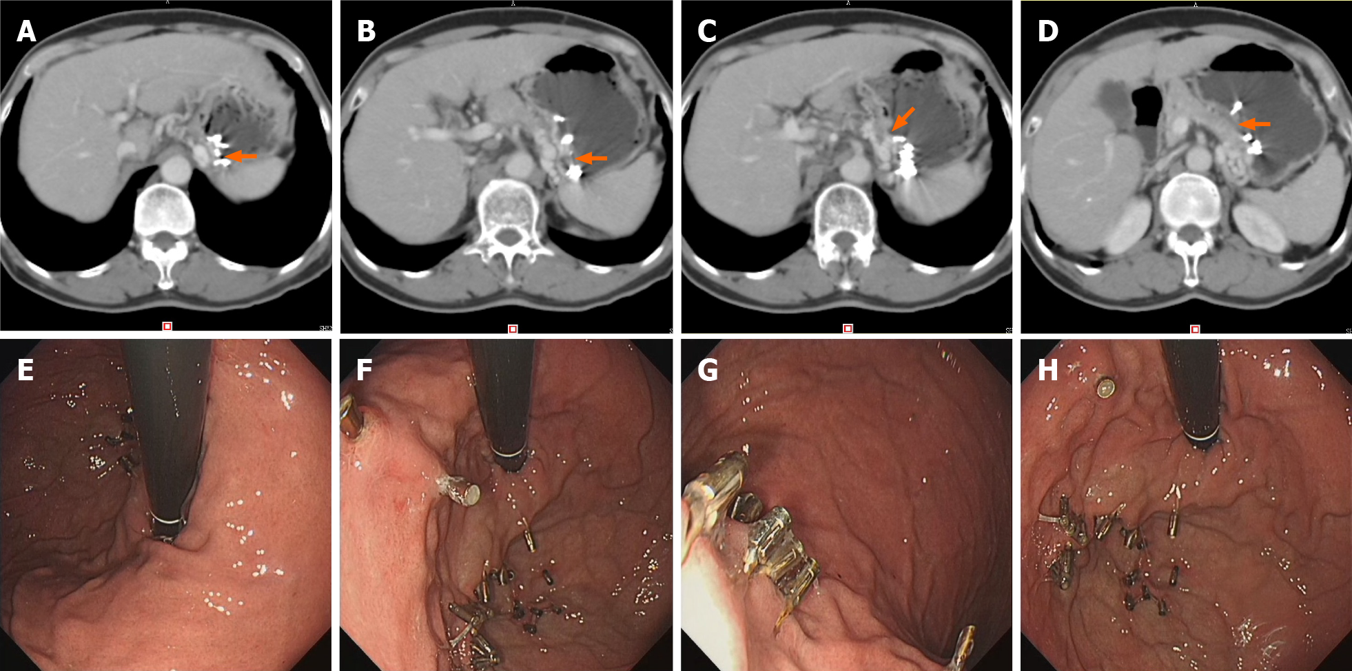
Figure 2 Follow up imaging and endoscopy images.
A-D: Imaging follow-up showing the significantly improved gastric varices (arrows) at the 5th postoperative month; E-H: Gastroscopy showing the clips still in place with well-healed varices at the 5th postoperative month.
- Citation: Yang GC, Mo YX, Zhang WH, Zhou LB, Huang XM, Cao LM. Endoscopic clipping for the secondary prophylaxis of bleeding gastric varices in a patient with cirrhosis: A case report. World J Clin Cases 2022; 10(4): 1447-1453
- URL: https://www.wjgnet.com/2307-8960/full/v10/i4/1447.htm
- DOI: https://dx.doi.org/10.12998/wjcc.v10.i4.1447


