Copyright
©The Author(s) 2022.
World J Clin Cases. Feb 6, 2022; 10(4): 1366-1372
Published online Feb 6, 2022. doi: 10.12998/wjcc.v10.i4.1366
Published online Feb 6, 2022. doi: 10.12998/wjcc.v10.i4.1366
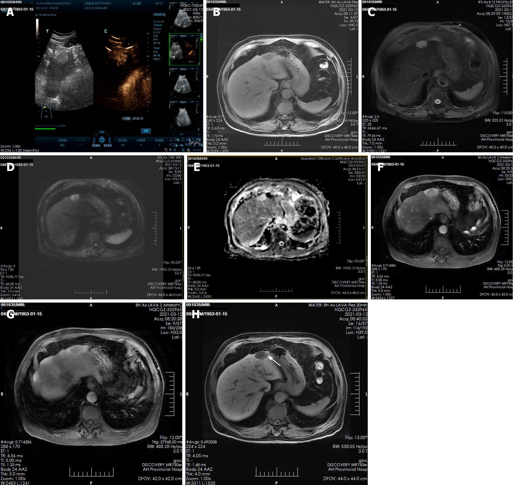
Figure 1 Imaging findings in a 68-year-old man with biliary adenofibroma.
A: Ultrasonography showed hyperechoic nodules in the left lobe of the liver, after injection of contrast agent, it showed early and obvious enhancement; B and C: Plain magnetic resonance imaging (MRI) scan showed hypointensity on T1-weighted imaging (T1WI) and hyperintensity on T2-weighted imaging (T2WI); D and E: Diffusion-weighted imaging (DWI) showed moderate hyperintensity and isointensity on ADC; F-H: Enhanced MRI scan showed obvious enhancement of solid components and separation of lesions in the arterial phase, the degree of lesion enhancement decreased in the portal phase, in the hepatobiliary phase, enhancement of the bile duct structure was found in the lesion.
- Citation: Li SP, Wang P, Deng KX. Imaging presentation of biliary adenofibroma: A case report. World J Clin Cases 2022; 10(4): 1366-1372
- URL: https://www.wjgnet.com/2307-8960/full/v10/i4/1366.htm
- DOI: https://dx.doi.org/10.12998/wjcc.v10.i4.1366


