Copyright
©The Author(s) 2022.
World J Clin Cases. Feb 6, 2022; 10(4): 1291-1295
Published online Feb 6, 2022. doi: 10.12998/wjcc.v10.i4.1291
Published online Feb 6, 2022. doi: 10.12998/wjcc.v10.i4.1291
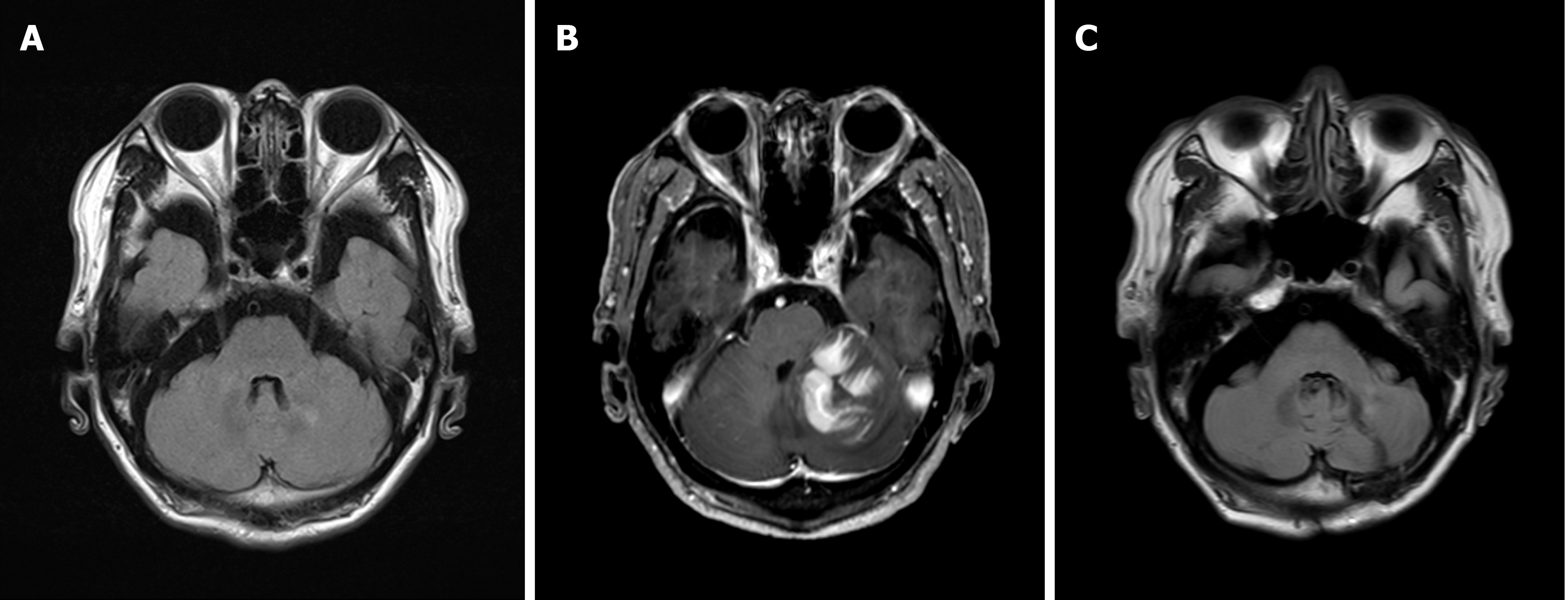
Figure 2 Findings of brain magnetic resonance imaging.
A: Image showing no evidence of space-occupying intracranial or diffusion-restricted lesions. The image was taken at the first clinical visit; B: Image showing an irregular contrast-enhancing lesion, measuring approximately 3.8 cm × 3.8 cm in size, with diffusion restriction in the left cerebellum. The image was taken 4 mon after the first clinical visit; C: Image showing no evidence of abnormal contrast-enhancing lesion in the cerebellum after six cycles of systemic chemotherapy.
- Citation: Jang HR, Lim KH, Lee K. Primary central nervous system lymphoma presenting as a single choroidal lesion mimicking metastasis: A case report. World J Clin Cases 2022; 10(4): 1291-1295
- URL: https://www.wjgnet.com/2307-8960/full/v10/i4/1291.htm
- DOI: https://dx.doi.org/10.12998/wjcc.v10.i4.1291


