Copyright
©The Author(s) 2022.
World J Clin Cases. Dec 26, 2022; 10(36): 13451-13457
Published online Dec 26, 2022. doi: 10.12998/wjcc.v10.i36.13451
Published online Dec 26, 2022. doi: 10.12998/wjcc.v10.i36.13451
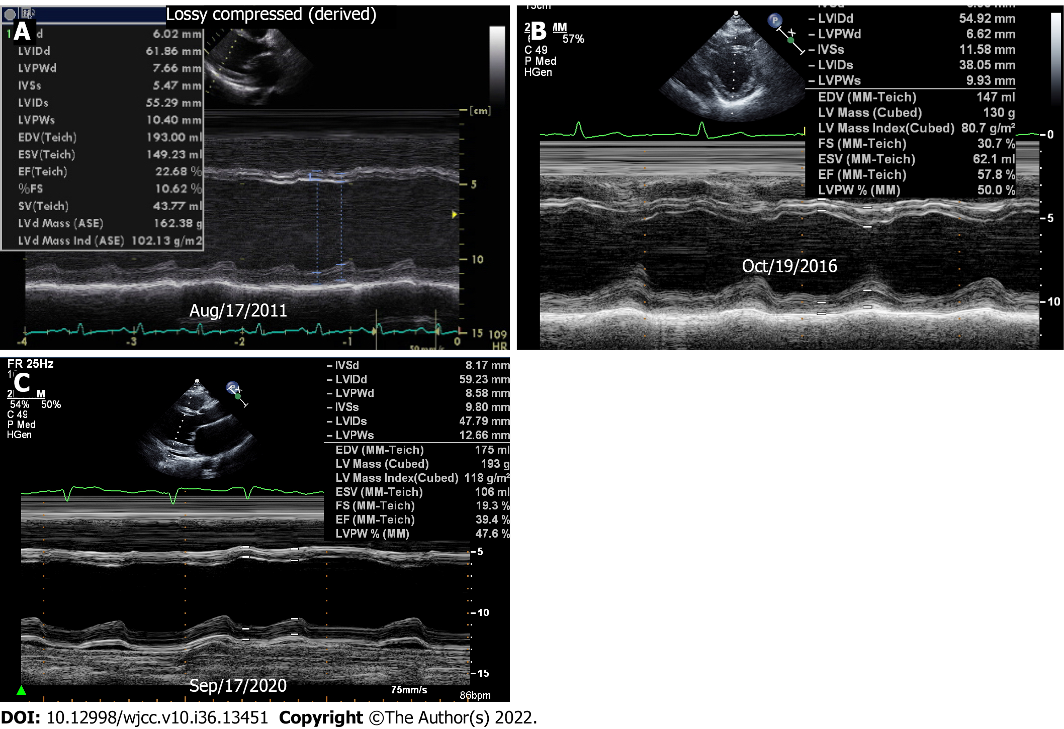
Figure 2 Transthoracic echocardiography.
A: Baseline echocardiography showed dilated left ventricular (LV) dimension and severely decreased LV ejection fraction (EF); B: Preserved LVEF at one year follow-up; C: The last M-mode image showed dilated LV and low LVEF again. Movies of the serial change of 4-chamber views showed the same findings.
- Citation: Lee SD, Lee HJ, Kim HR, Kang MG, Kim K, Park JR. Development of dilated cardiomyopathy with a long latent period followed by viral fulminant myocarditis: A case report. World J Clin Cases 2022; 10(36): 13451-13457
- URL: https://www.wjgnet.com/2307-8960/full/v10/i36/13451.htm
- DOI: https://dx.doi.org/10.12998/wjcc.v10.i36.13451


