Copyright
©The Author(s) 2022.
World J Clin Cases. Dec 26, 2022; 10(36): 13418-13425
Published online Dec 26, 2022. doi: 10.12998/wjcc.v10.i36.13418
Published online Dec 26, 2022. doi: 10.12998/wjcc.v10.i36.13418
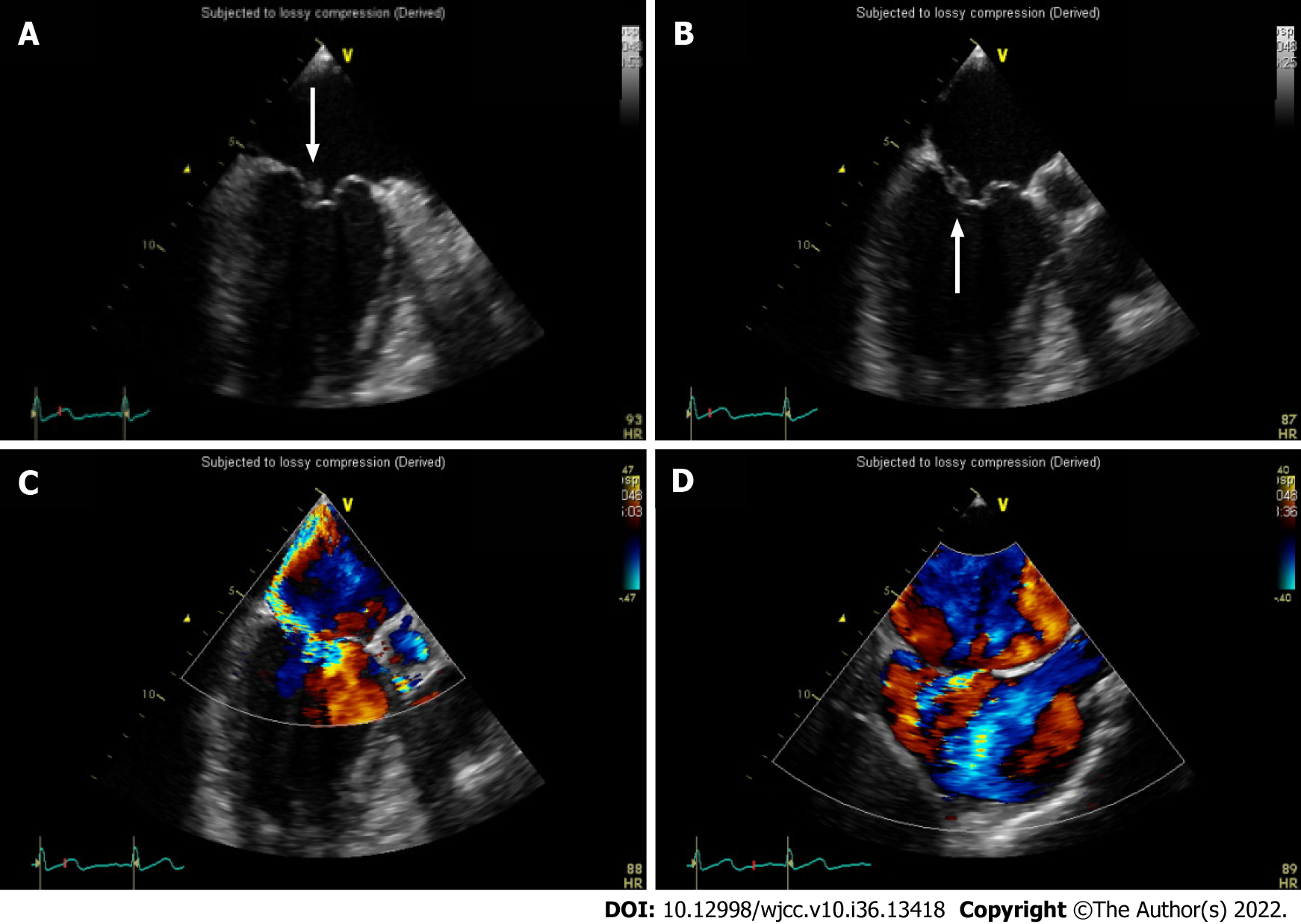
Figure 3 Transesophageal echocardiography.
A and B: The anterior leaflet of the mitral valve was detected with a wart (white arrow) of a cord-like medium echoic substance about 10mm and the posterior leaflet was detected with the wart (white arrow) of a medium echoic substance about 7 mm × 7 mm; C and D: There was severe mitral regurgitation and the regurgitation bundle was distributed along the posterior leaflet of the mitral valve. There was mild tricuspid valve and the aortic valve regurgitation.
- Citation: Chen Y, Chen D, Liu H, Zhang CG, Song LL. Staphylococcus aureus bacteremia and infective endocarditis in a patient with epidermolytic hyperkeratosis: A case report. World J Clin Cases 2022; 10(36): 13418-13425
- URL: https://www.wjgnet.com/2307-8960/full/v10/i36/13418.htm
- DOI: https://dx.doi.org/10.12998/wjcc.v10.i36.13418


