Copyright
©The Author(s) 2022.
World J Clin Cases. Dec 26, 2022; 10(36): 13418-13425
Published online Dec 26, 2022. doi: 10.12998/wjcc.v10.i36.13418
Published online Dec 26, 2022. doi: 10.12998/wjcc.v10.i36.13418
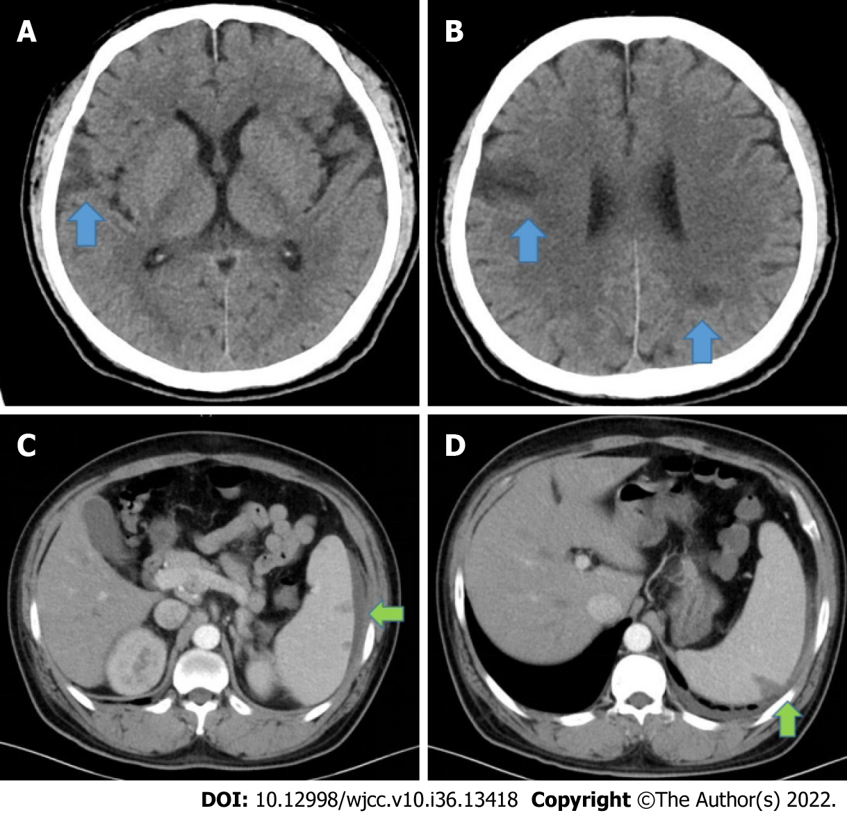
Figure 2 Computed tomography.
A and B: Cranial computed tomography showing multiple low-density shadows (blue arrow, A and B) in the brain; C and D: Abdominal enhanced CT scan showing multiple low-density shadows (green arrow, C and D) in the spleen, considered a splenic abscess with subcapsular effusion.
- Citation: Chen Y, Chen D, Liu H, Zhang CG, Song LL. Staphylococcus aureus bacteremia and infective endocarditis in a patient with epidermolytic hyperkeratosis: A case report. World J Clin Cases 2022; 10(36): 13418-13425
- URL: https://www.wjgnet.com/2307-8960/full/v10/i36/13418.htm
- DOI: https://dx.doi.org/10.12998/wjcc.v10.i36.13418


