
















©The Author(s) 2022.
World J Clin Cases. Dec 16, 2022; 10(35): 13088-13098
Published online Dec 16, 2022. doi: 10.12998/wjcc.v10.i35.13088
Published online Dec 16, 2022. doi: 10.12998/wjcc.v10.i35.13088
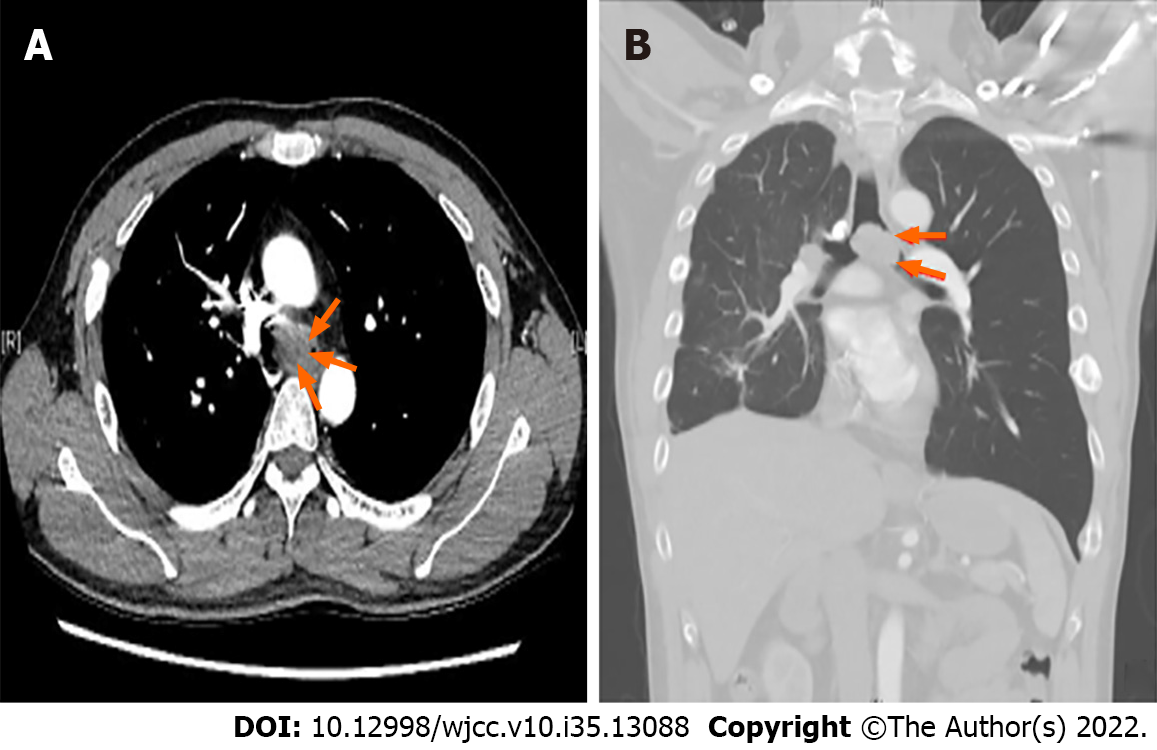
Figure 1 Preoperative chest computed tomography images of patient.
A: Axial view; B: Coronal view. These computed tomography images show a large tracheal protruding mass (orange arrow) at the carinal level, approximately 3.0 cm in size, causing nearly total obstruction of the left main bronchus and left lung hyperinflation.
- Citation: Liu IL, Chou AH, Chiu CH, Cheng YT, Lin HT. Tracheostomy and venovenous extracorporeal membrane oxygenation for difficult airway patient with carinal melanoma: A case report and literature review. World J Clin Cases 2022; 10(35): 13088-13098
- URL: https://www.wjgnet.com/2307-8960/full/v10/i35/13088.htm
- DOI: https://dx.doi.org/10.12998/wjcc.v10.i35.13088