Copyright
©The Author(s) 2022.
World J Clin Cases. Dec 16, 2022; 10(35): 12854-12874
Published online Dec 16, 2022. doi: 10.12998/wjcc.v10.i35.12854
Published online Dec 16, 2022. doi: 10.12998/wjcc.v10.i35.12854
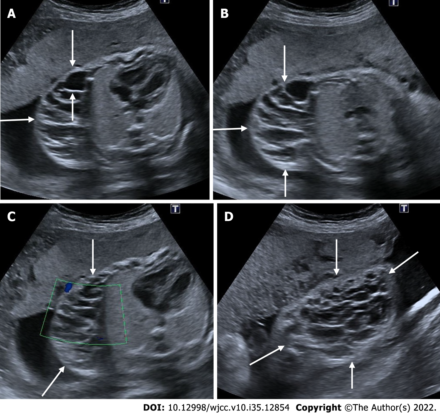
Figure 25 Cystic lymphangioma.
24 wk of gestation. A and B: On ultrasonography, a complex cystic heterogeneous lesion with multicystic areas on the right side of the fetal thoracic wall, under the skin (arrows); C: There was seen no vascularity on Doppler sonography; D: There is a view of the cystic lesion (arrows) on the chest wall from a different section.
- Citation: Ece B, Aydın S, Kantarci M. Antenatal imaging: A pictorial review. World J Clin Cases 2022; 10(35): 12854-12874
- URL: https://www.wjgnet.com/2307-8960/full/v10/i35/12854.htm
- DOI: https://dx.doi.org/10.12998/wjcc.v10.i35.12854


