Copyright
©The Author(s) 2022.
World J Clin Cases. Dec 16, 2022; 10(35): 12854-12874
Published online Dec 16, 2022. doi: 10.12998/wjcc.v10.i35.12854
Published online Dec 16, 2022. doi: 10.12998/wjcc.v10.i35.12854
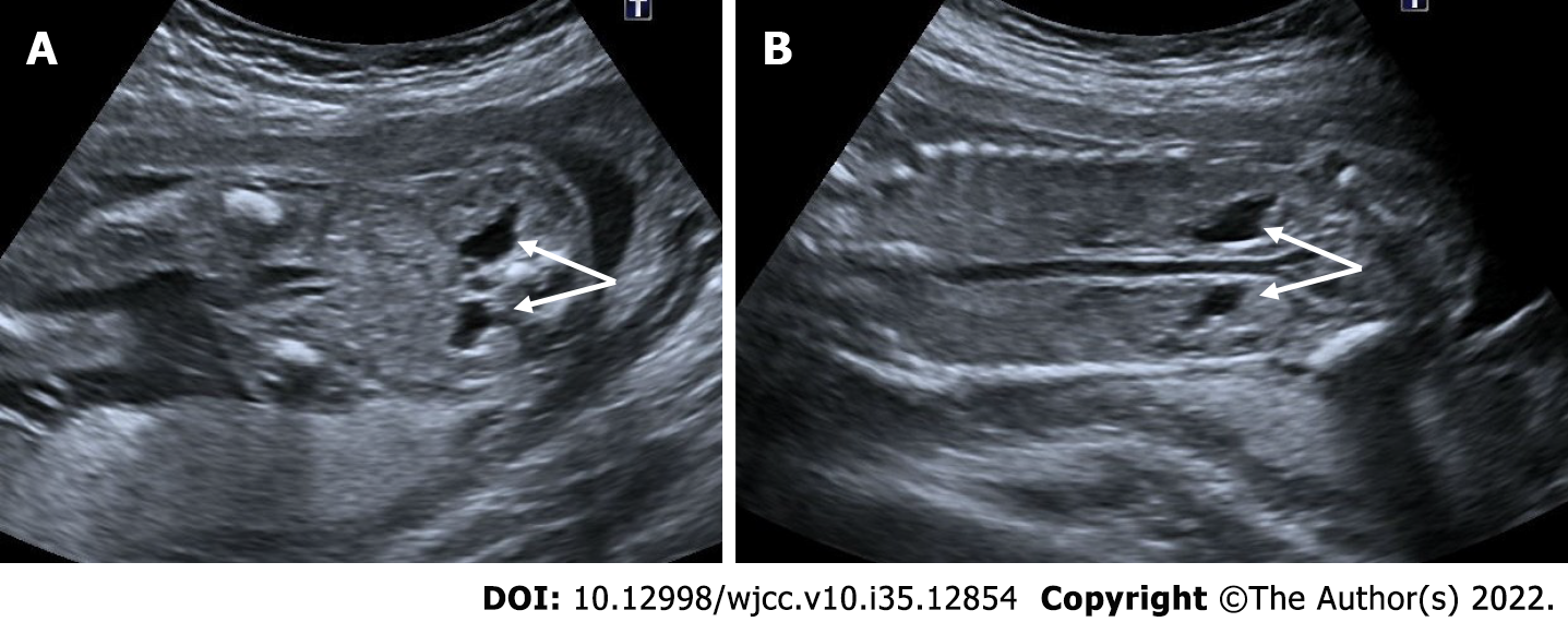
Figure 20 Bilateral mild hydronephrosis.
A and B: 23 wk of gestation. On ultrasonography, bilateral renal pelvis was wider than normal (arrows). Renal pelvis diameters were 6 mm on the right and 5 mm on the left. There was no dilatation of the bilateral ureters.
- Citation: Ece B, Aydın S, Kantarci M. Antenatal imaging: A pictorial review. World J Clin Cases 2022; 10(35): 12854-12874
- URL: https://www.wjgnet.com/2307-8960/full/v10/i35/12854.htm
- DOI: https://dx.doi.org/10.12998/wjcc.v10.i35.12854


