Copyright
©The Author(s) 2022.
World J Clin Cases. Dec 16, 2022; 10(35): 12854-12874
Published online Dec 16, 2022. doi: 10.12998/wjcc.v10.i35.12854
Published online Dec 16, 2022. doi: 10.12998/wjcc.v10.i35.12854
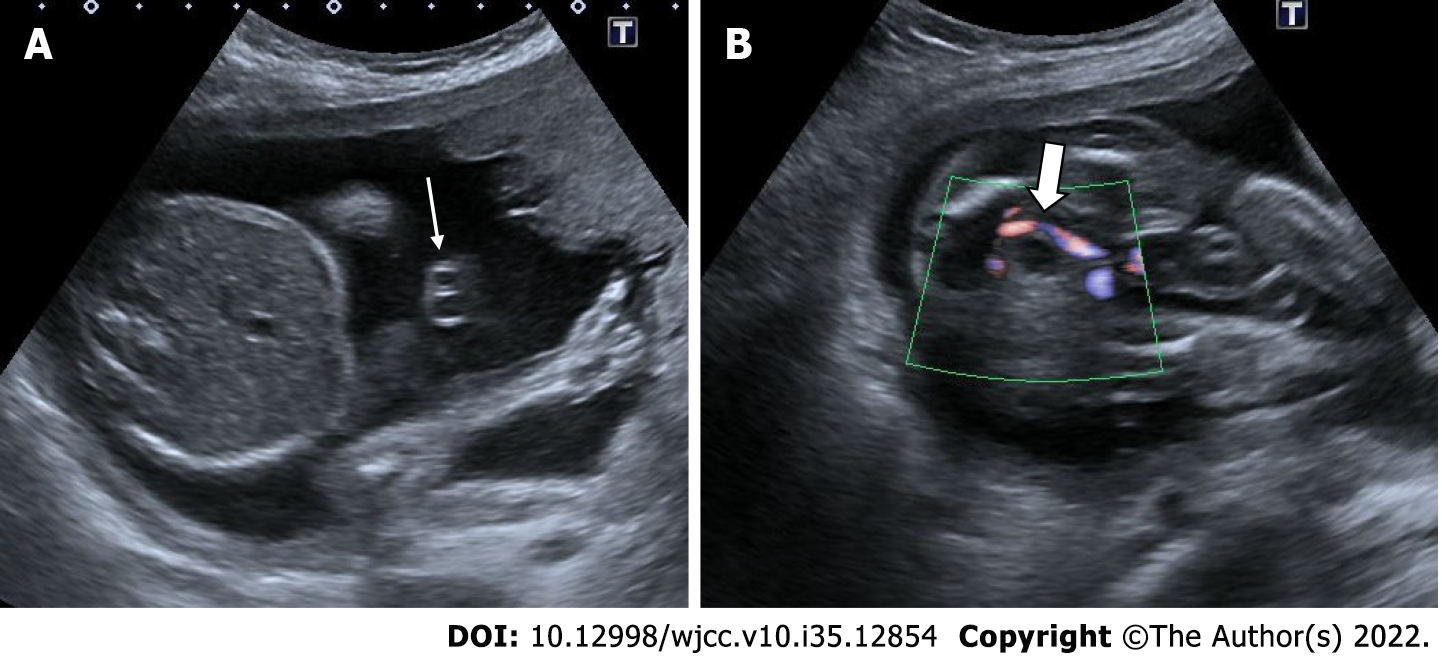
Figure 10 Single umbilical artery.
29-years-old, first pregnancy, 21 wk of gestation. A: On ultrasonography, there was one artery (thin arrow) and one vein in the umbilical cord; B: Doppler ultrasonography reveals a solitary artery at the level of the bladder in the fetal pelvis (thick arrow).
- Citation: Ece B, Aydın S, Kantarci M. Antenatal imaging: A pictorial review. World J Clin Cases 2022; 10(35): 12854-12874
- URL: https://www.wjgnet.com/2307-8960/full/v10/i35/12854.htm
- DOI: https://dx.doi.org/10.12998/wjcc.v10.i35.12854


