Copyright
©The Author(s) 2022.
World J Clin Cases. Dec 6, 2022; 10(34): 12781-12786
Published online Dec 6, 2022. doi: 10.12998/wjcc.v10.i34.12781
Published online Dec 6, 2022. doi: 10.12998/wjcc.v10.i34.12781
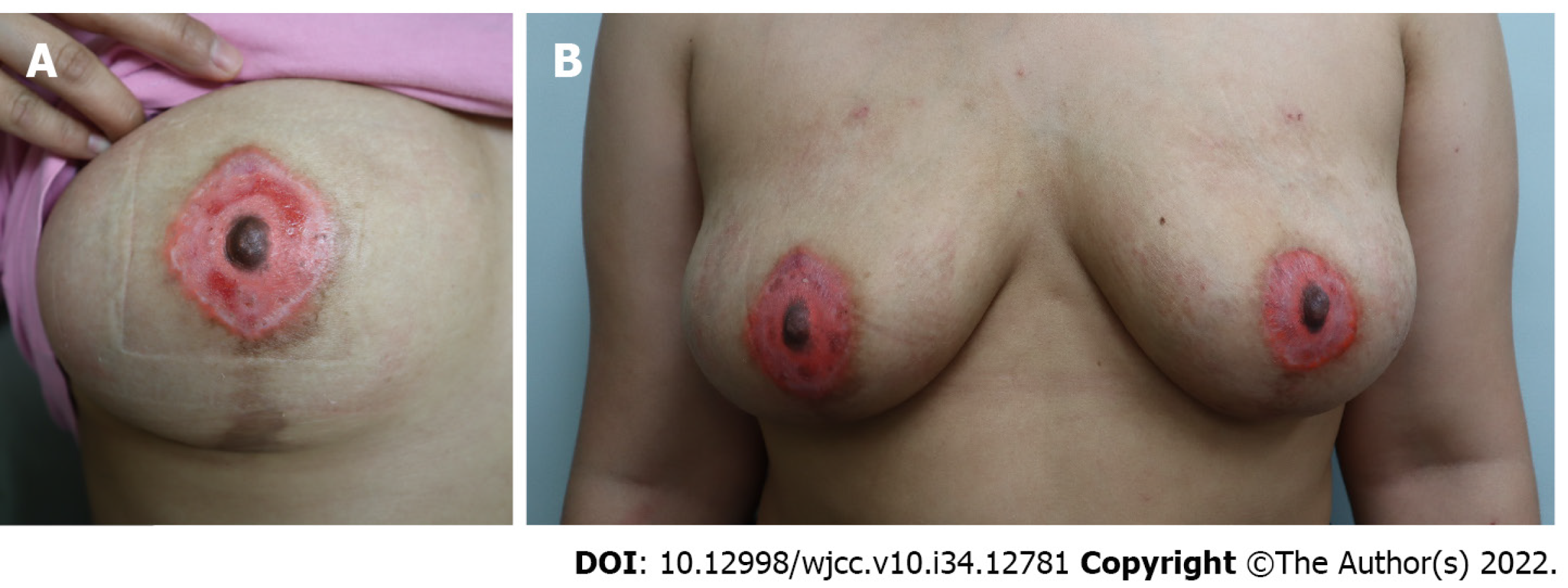
Figure 3 A photograph taken after treatment.
A: A photograph taken after two weeks of treatment. Swelling, discharge, and pain improved, and bleeding stopped. Epithelialization was in progress, but there was still a raw surface on the areolar area. Hyperpigmentation and hypopigmentation were seen. Also, color mismatch and size mismatch of both nipple areolar complex (NACs) were noted, and there was a color mismatch between the nipple and areolar area, which looked unnatural; B: A photograph taken after four weeks of treatment. After treatment, more than half of the ink was lost, leaving only a part. Hyperpigmentation and hypopigmentation had worsened. It was necessary to correct the size and color of both NACs. The areolar tissue had turned into scar tissue and looked even more unnatural.
- Citation: Byeon JY, Kim TH, Choi HJ. Complication after nipple-areolar complex tattooing performed by a non-medical person: A case report. World J Clin Cases 2022; 10(34): 12781-12786
- URL: https://www.wjgnet.com/2307-8960/full/v10/i34/12781.htm
- DOI: https://dx.doi.org/10.12998/wjcc.v10.i34.12781


