Copyright
©The Author(s) 2022.
World J Clin Cases. Dec 6, 2022; 10(34): 12768-12774
Published online Dec 6, 2022. doi: 10.12998/wjcc.v10.i34.12768
Published online Dec 6, 2022. doi: 10.12998/wjcc.v10.i34.12768
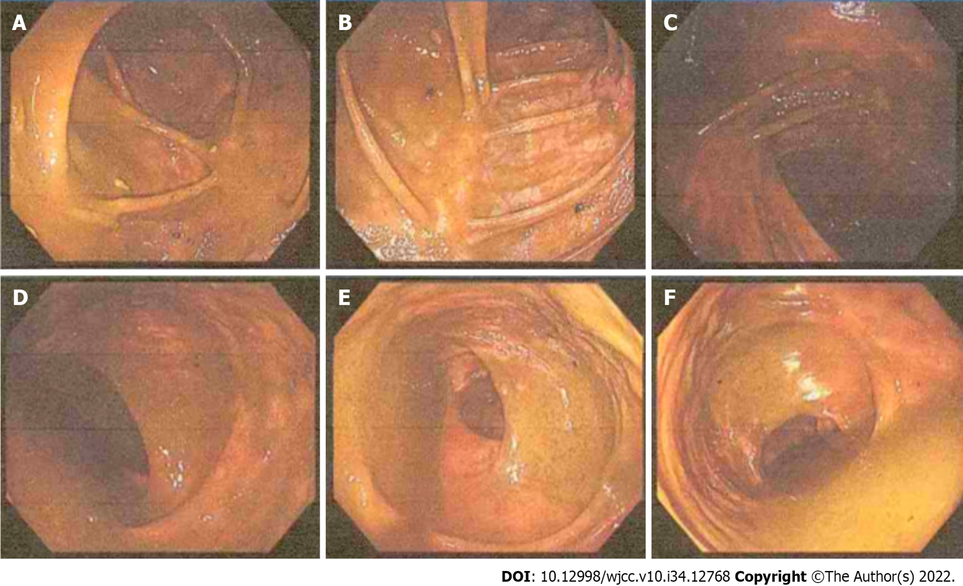
Figure 2 Colonoscopy images show a large amount of fecal residue in the intestinal lumen, which was dilated (dominated by the dilation of the ascending colon, hepatic region, and transverse colon), and a smooth mucosa.
No obvious structural abnormalities were found. A: Ileocecum; B: Ascending colon proximal to the liver; C: Transverse colon proximal to the liver; D: Descending colon; E: Sigmoid colon; F: Rectum.
- Citation: Zhang ZM, Kong S, Gao XX, Jia XH, Zheng CN. Colonic tubular duplication combined with congenital megacolon: A case report. World J Clin Cases 2022; 10(34): 12768-12774
- URL: https://www.wjgnet.com/2307-8960/full/v10/i34/12768.htm
- DOI: https://dx.doi.org/10.12998/wjcc.v10.i34.12768


