Copyright
©The Author(s) 2022.
World J Clin Cases. Dec 6, 2022; 10(34): 12711-12716
Published online Dec 6, 2022. doi: 10.12998/wjcc.v10.i34.12711
Published online Dec 6, 2022. doi: 10.12998/wjcc.v10.i34.12711
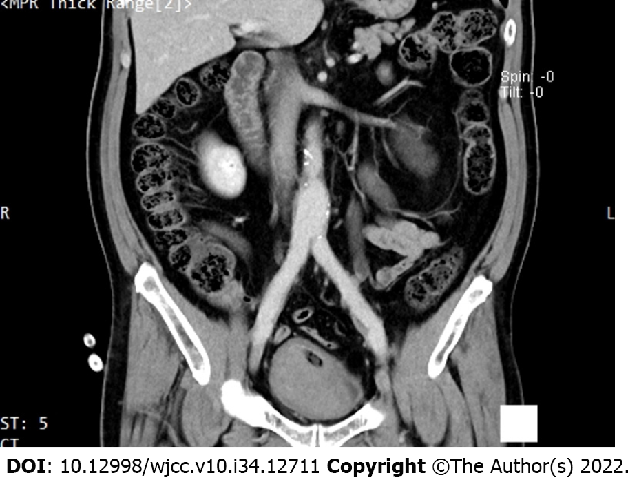
Figure 1 Contrast-enhanced computed tomography of the urinary system, and computed tomography angiography examination showed that the left renal pelvis and left ureter were dilated, and patchy high-density shadow could be seen inside.
- Citation: Feng T, Zhao X, Zhu L, Chen W, Gao YL, Wei JL. Ureteral- artificial iliac artery fistula: A case report. World J Clin Cases 2022; 10(34): 12711-12716
- URL: https://www.wjgnet.com/2307-8960/full/v10/i34/12711.htm
- DOI: https://dx.doi.org/10.12998/wjcc.v10.i34.12711


