Copyright
©The Author(s) 2022.
World J Clin Cases. Nov 26, 2022; 10(33): 12447-12454
Published online Nov 26, 2022. doi: 10.12998/wjcc.v10.i33.12447
Published online Nov 26, 2022. doi: 10.12998/wjcc.v10.i33.12447
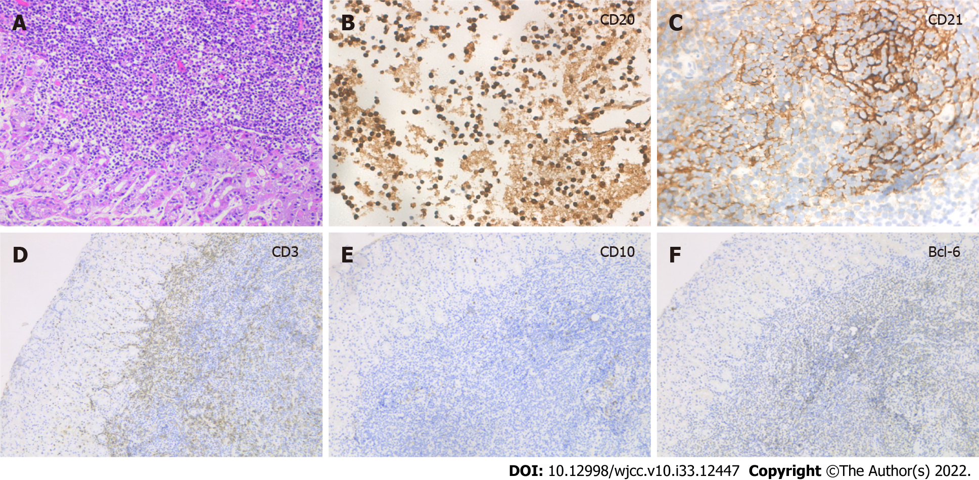
Figure 2 Immunohistochemical results of the gastric mucosa-associated lymphoid tissue lymphoma.
A: Hematoxylin-eosin staining of extensive lymphocytic infiltration (200×); B: Immunohistochemistry showed that the lymphoid cells were diffusely positive for CD20 (400×); C: CD21 showed expansion and destruction of follicular dendritic cells (400×); D: Immunohistochemical stains showed CD3 negative (100×); E: CD10 was negative (100×); F: Bcl-6 was negative (100×).
- Citation: Lu SN, Huang C, Li LL, Di LJ, Yao J, Tuo BG, Xie R. Synchronous early gastric and intestinal mucosa-associated lymphoid tissue lymphoma in a Helicobacter pylori-negative patient: A case report. World J Clin Cases 2022; 10(33): 12447-12454
- URL: https://www.wjgnet.com/2307-8960/full/v10/i33/12447.htm
- DOI: https://dx.doi.org/10.12998/wjcc.v10.i33.12447


