Copyright
©The Author(s) 2022.
World J Clin Cases. Nov 26, 2022; 10(33): 12447-12454
Published online Nov 26, 2022. doi: 10.12998/wjcc.v10.i33.12447
Published online Nov 26, 2022. doi: 10.12998/wjcc.v10.i33.12447
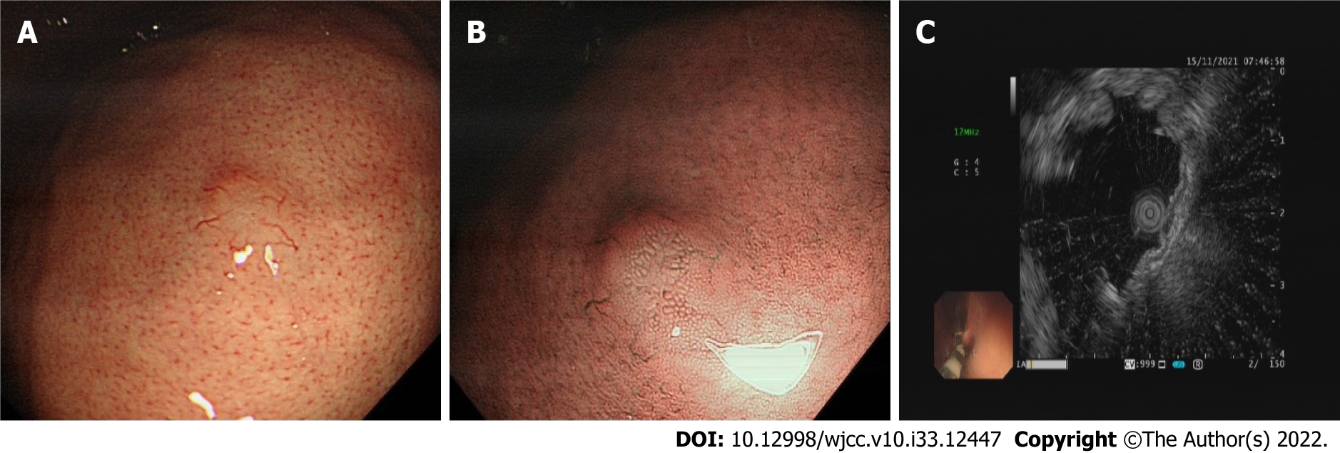
Figure 1 Endoscopic images of the gastric lesion.
A: A solitary submucosal eminence was observed; B: Narrow-band imaging appearance of gastric pit pattern elongates and expands, with appearance of irregular abnormal vessels; C: Endoscopic ultrasonography showed thickening of muscularis mucosa with a hypoechoic lesion 2 mm × 5 mm in size.
- Citation: Lu SN, Huang C, Li LL, Di LJ, Yao J, Tuo BG, Xie R. Synchronous early gastric and intestinal mucosa-associated lymphoid tissue lymphoma in a Helicobacter pylori-negative patient: A case report. World J Clin Cases 2022; 10(33): 12447-12454
- URL: https://www.wjgnet.com/2307-8960/full/v10/i33/12447.htm
- DOI: https://dx.doi.org/10.12998/wjcc.v10.i33.12447


