Copyright
©The Author(s) 2022.
World J Clin Cases. Nov 26, 2022; 10(33): 12313-12318
Published online Nov 26, 2022. doi: 10.12998/wjcc.v10.i33.12313
Published online Nov 26, 2022. doi: 10.12998/wjcc.v10.i33.12313
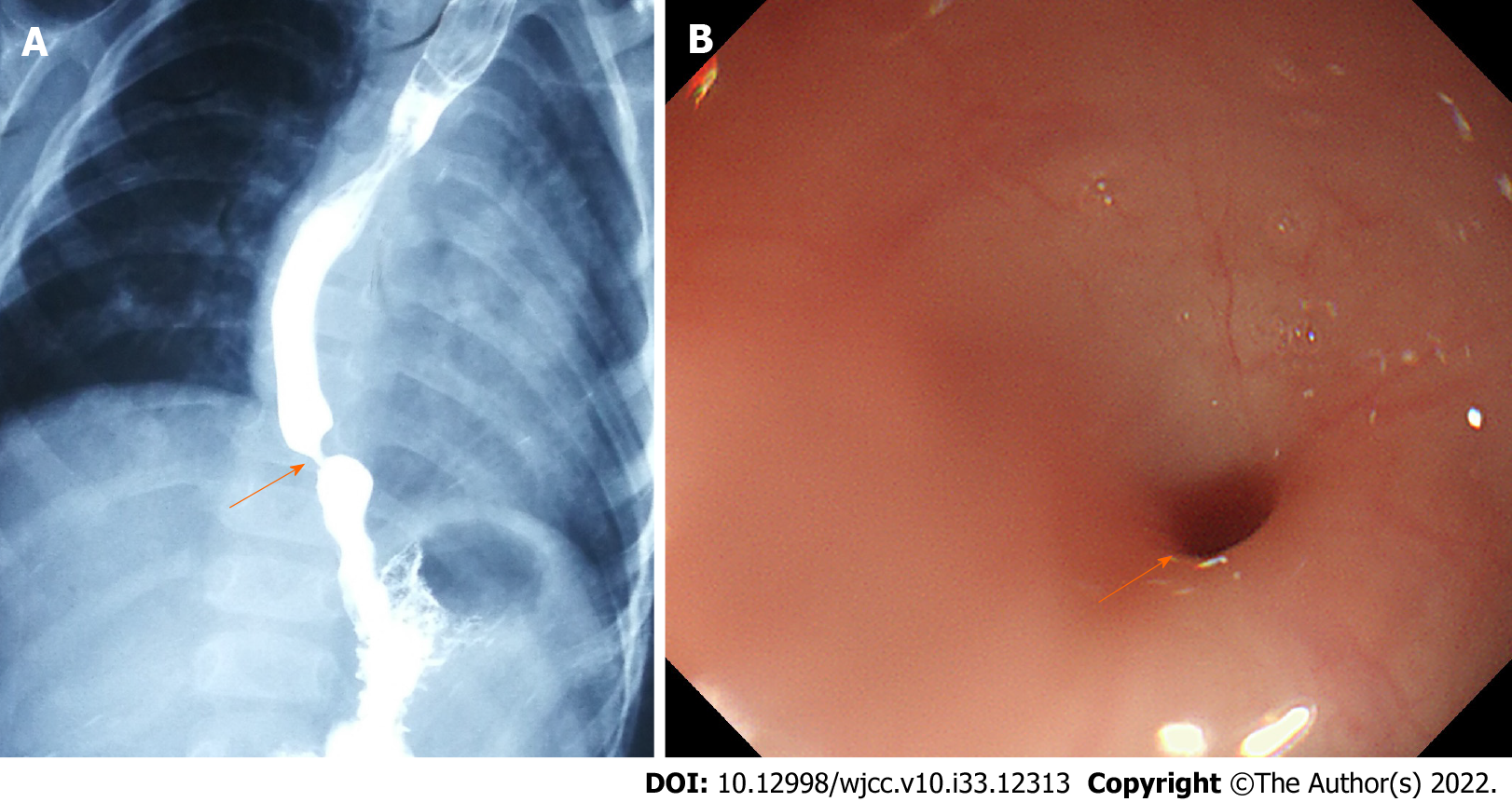
Figure 1 Esophagography and gastroendoscopy of the esophagus.
A: Esophagography confirmed that the stenosis was located in the lower esophagus (arrow); B: The congenital stenosis opening in the esophagus with a < 3 mm inner diameter was observed by gastroendoscopy (arrow).
- Citation: Liu SQ, Lv Y, Luo RX. Endoscopic magnetic compression stricturoplasty for congenital esophageal stenosis: A case report. World J Clin Cases 2022; 10(33): 12313-12318
- URL: https://www.wjgnet.com/2307-8960/full/v10/i33/12313.htm
- DOI: https://dx.doi.org/10.12998/wjcc.v10.i33.12313


