Copyright
©The Author(s) 2022.
World J Clin Cases. Nov 16, 2022; 10(32): 12028-12035
Published online Nov 16, 2022. doi: 10.12998/wjcc.v10.i32.12028
Published online Nov 16, 2022. doi: 10.12998/wjcc.v10.i32.12028
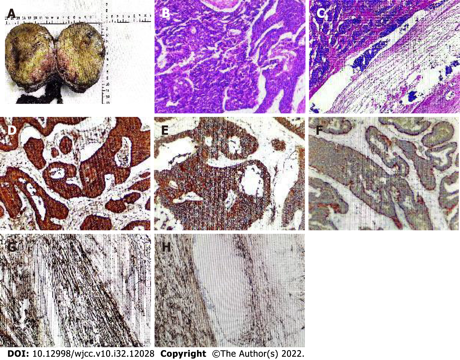
Figure 3 Postoperative pathology and immunohistochemistry.
A: The resected specimen consisted of the right testis, tumor, spermatic cord, and epididymis; B and C: Histological HE staining of primary neuroendocrine tumors of the testis; D: Tumor cells positive for CgA; E: Tumor cells positive for CD56; F: Syn-positive tumor cells; G and H: Tumor cells CD34 negative.
- Citation: Xiao T, Luo LH, Guo LF, Wang LQ, Feng L. Primary testicular neuroendocrine tumor with liver lymph node metastasis: A case report and review of the literature. World J Clin Cases 2022; 10(32): 12028-12035
- URL: https://www.wjgnet.com/2307-8960/full/v10/i32/12028.htm
- DOI: https://dx.doi.org/10.12998/wjcc.v10.i32.12028


