Copyright
©The Author(s) 2022.
World J Clin Cases. Nov 16, 2022; 10(32): 11929-11935
Published online Nov 16, 2022. doi: 10.12998/wjcc.v10.i32.11929
Published online Nov 16, 2022. doi: 10.12998/wjcc.v10.i32.11929
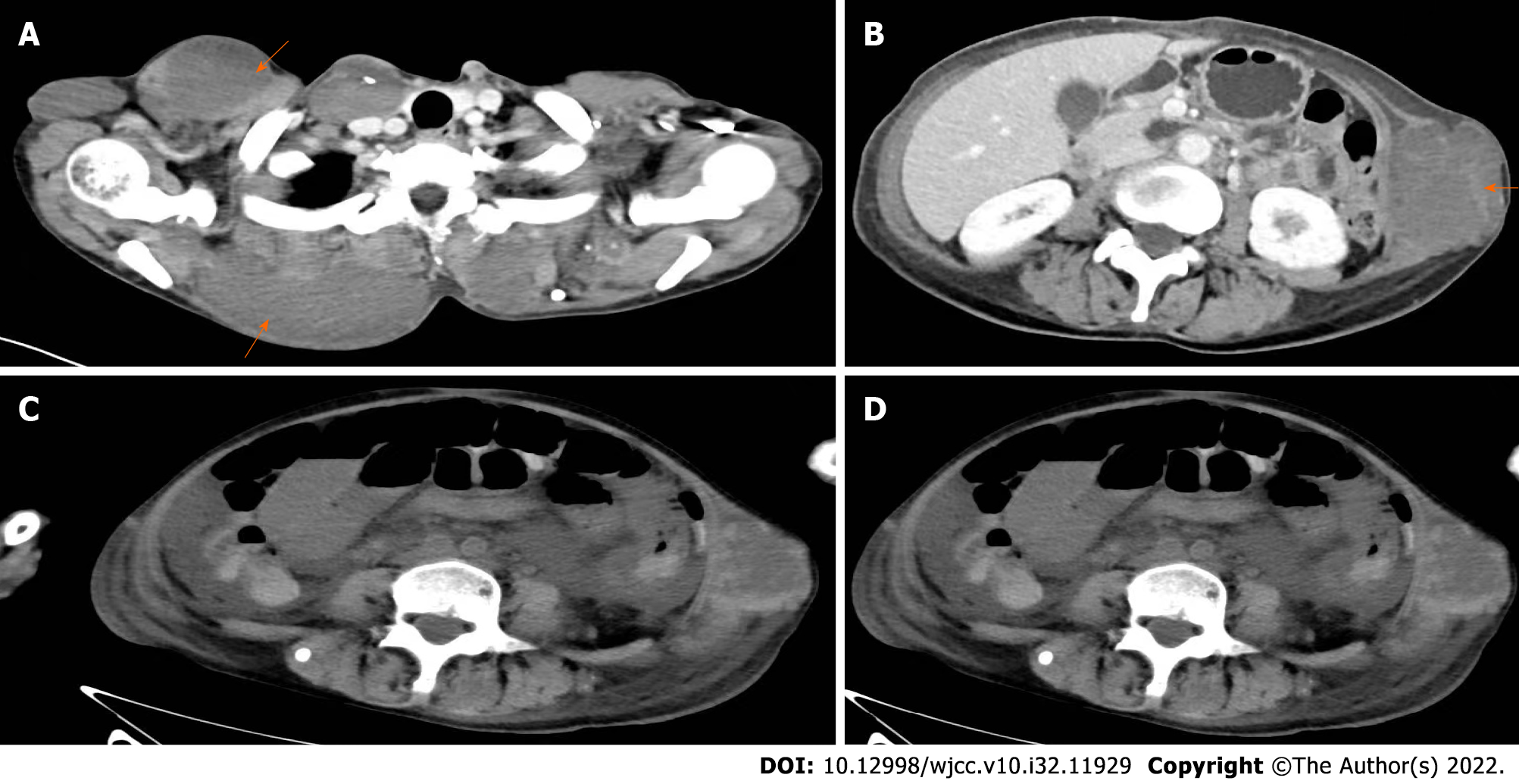
Figure 2 Computed tomography.
A: Hemangioma on the front and back of the abdomen (orange arrows); B: Hemangioma on the left side of the abdomen; C and D: Representative abdominal scans showing an obstruction in the small intestine.
- Citation: Zhai JH, Li SX, Jin G, Zhang YY, Zhong WL, Chai YF, Wang BM. Blue rubber bleb nevus syndrome complicated with disseminated intravascular coagulation and intestinal obstruction: A case report. World J Clin Cases 2022; 10(32): 11929-11935
- URL: https://www.wjgnet.com/2307-8960/full/v10/i32/11929.htm
- DOI: https://dx.doi.org/10.12998/wjcc.v10.i32.11929


