Copyright
©The Author(s) 2022.
World J Clin Cases. Nov 16, 2022; 10(32): 11827-11834
Published online Nov 16, 2022. doi: 10.12998/wjcc.v10.i32.11827
Published online Nov 16, 2022. doi: 10.12998/wjcc.v10.i32.11827
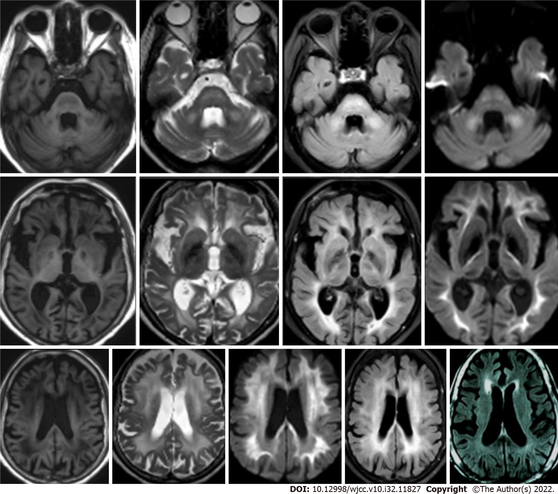
Figure 1 T1MRI, T2MRI, diffusion weighted image, and fluid-attenuated inversion recovery imaging of the brain.
All imaging indicated symmetrical, bilateral, patchy, and abnormal signals in the cerebral hemisphere, brain stem and cerebellar peduncle. Two years later, brain magnetic resonance imaging fluid-attenuated inversion recovery imaging showed that the white matter lesions were alleviated, and evidence of brain atrophy was observed.
- Citation: Zhao DH, Li QJ. Paraneoplastic neurological syndrome caused by cystitis glandularis: A case report and literature review. World J Clin Cases 2022; 10(32): 11827-11834
- URL: https://www.wjgnet.com/2307-8960/full/v10/i32/11827.htm
- DOI: https://dx.doi.org/10.12998/wjcc.v10.i32.11827


