
















©The Author(s) 2022.
World J Clin Cases. Nov 6, 2022; 10(31): 11646-11651
Published online Nov 6, 2022. doi: 10.12998/wjcc.v10.i31.11646
Published online Nov 6, 2022. doi: 10.12998/wjcc.v10.i31.11646
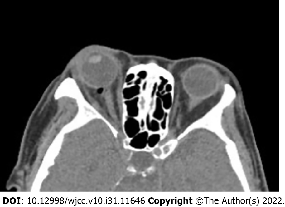
Figure 1 Postoperative head computed tomography.
Two hours after retrobulbar anesthesia, head computed tomography showed bubbles at the posterior of the patient’s right eye, partially compressing the optic nerve.
- Citation: Wang YL, Lan GR, Zou X, Wang EQ, Dai RP, Chen YX. Apnea caused by retrobulbar anesthesia: A case report. World J Clin Cases 2022; 10(31): 11646-11651
- URL: https://www.wjgnet.com/2307-8960/full/v10/i31/11646.htm
- DOI: https://dx.doi.org/10.12998/wjcc.v10.i31.11646