Copyright
©The Author(s) 2022.
World J Clin Cases. Nov 6, 2022; 10(31): 11549-11554
Published online Nov 6, 2022. doi: 10.12998/wjcc.v10.i31.11549
Published online Nov 6, 2022. doi: 10.12998/wjcc.v10.i31.11549
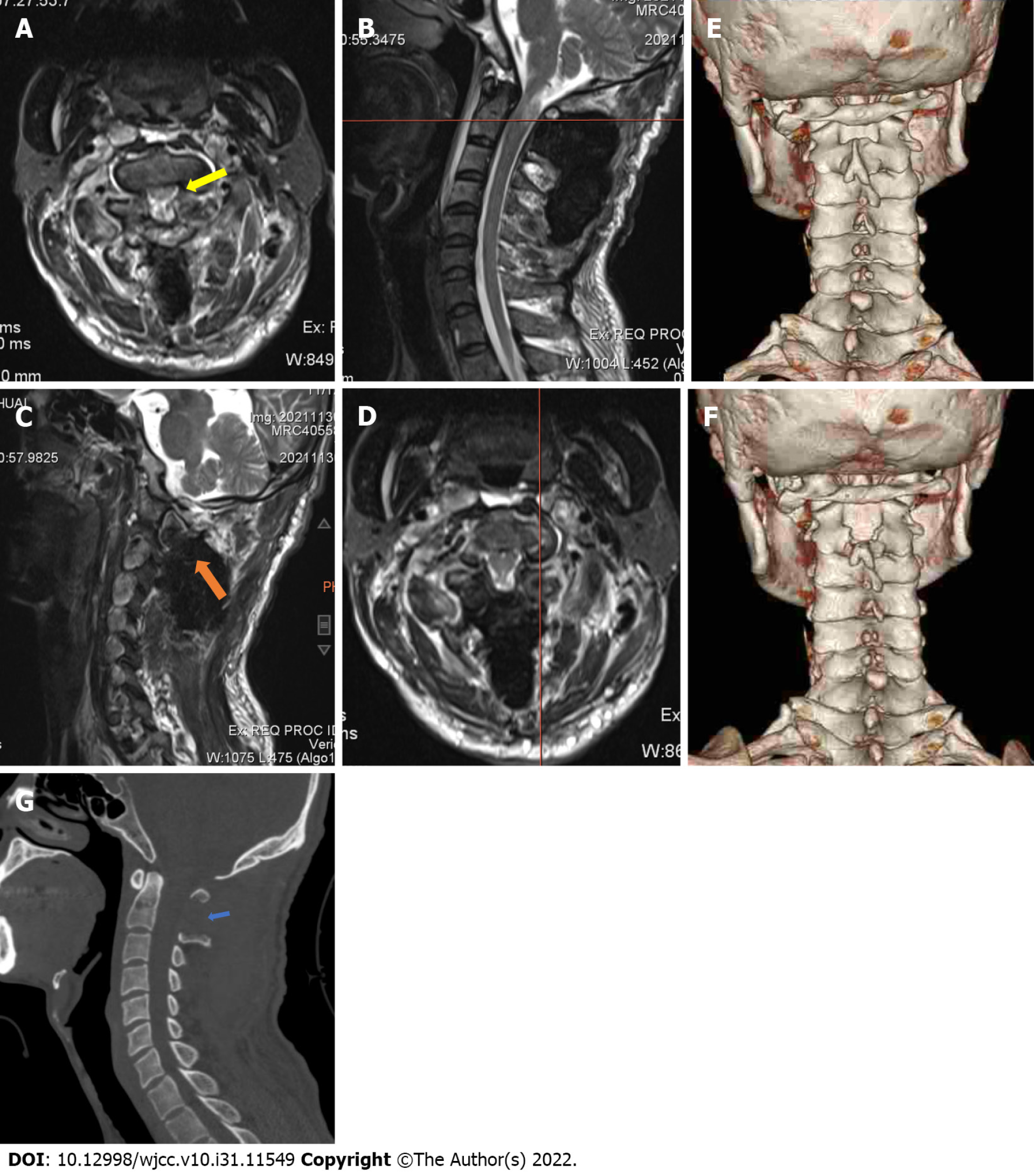
Figure 2 Post-operative images of the patient.
A: The coronal plane showed complete removal of the neoplasm and relief from compression of the spinal cord (yellow arrow) at the C1 position line (B); C: The sagittal plane also showed removal of the neoplasm (orange arrow) at the position line (D); Comparison of the pre-operative (E) and post-operative (F) images to three dimensional computed tomography showed limited structural changes on the posterior arch and laminar of the axis, with (G) substantial preservation of the upper cervical spine.
- Citation: Wang S, Ma JX, Zheng L, Sun ST, Xiang LB, Chen Y. Multiple bilateral and symmetric C1-2 ganglioneuromas: A case report. World J Clin Cases 2022; 10(31): 11549-11554
- URL: https://www.wjgnet.com/2307-8960/full/v10/i31/11549.htm
- DOI: https://dx.doi.org/10.12998/wjcc.v10.i31.11549


