Copyright
©The Author(s) 2022.
World J Clin Cases. Nov 6, 2022; 10(31): 11517-11522
Published online Nov 6, 2022. doi: 10.12998/wjcc.v10.i31.11517
Published online Nov 6, 2022. doi: 10.12998/wjcc.v10.i31.11517
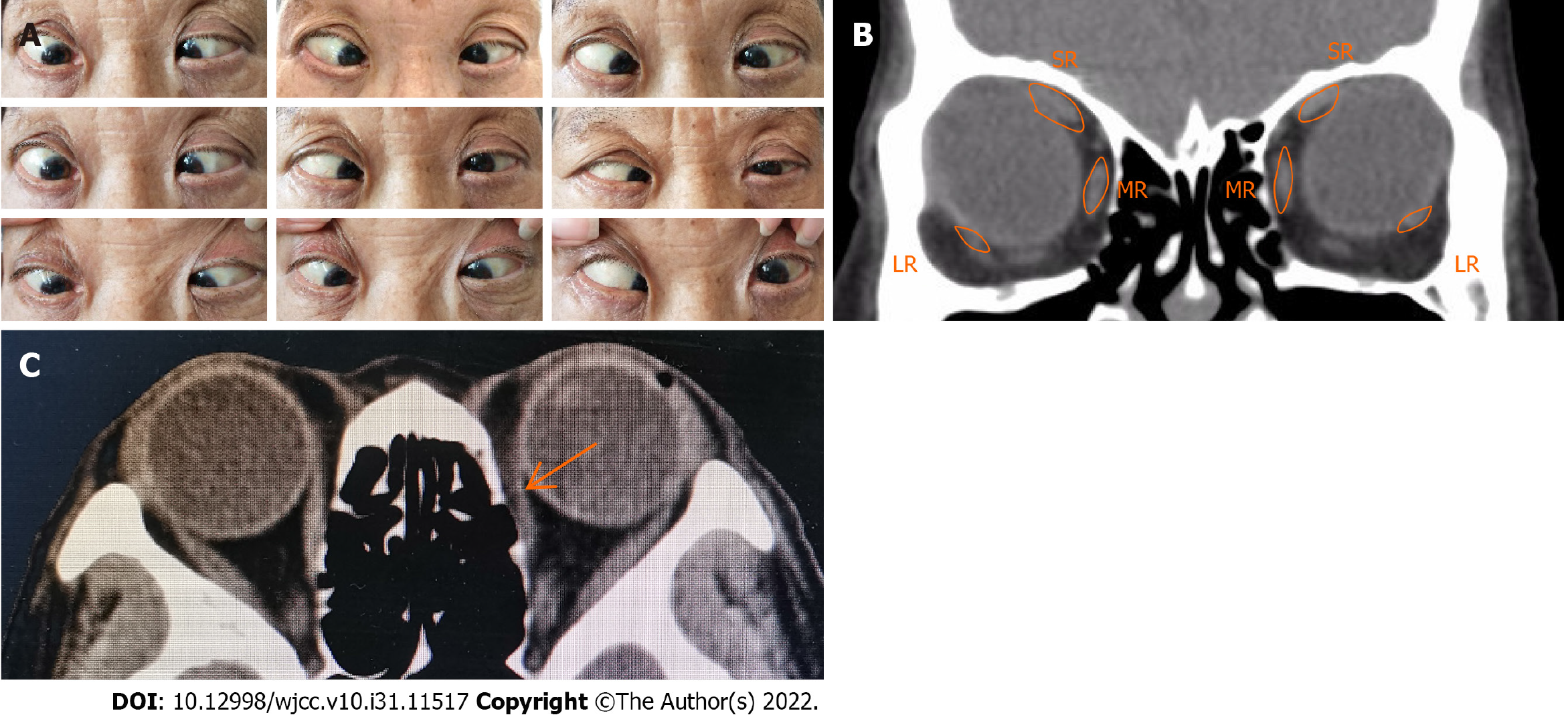
Figure 2 Preoperative clinical photographs.
A: Preoperative photograph showing the esotropia and hypotropia and the limitation of abduction and elevation in both eyes; B: Orbital computed tomography (CT) showing the displaced lateral rectus muscle inferiorly and superior rectos muscle medially; C: Orbital CT showing a suspected reattachment of the left medial rectus muscle to the globe (arrows). R: Superior rectus; LR: Lateral rectus.
- Citation: Yao Z, Jiang WL, Yang X. Yokoyama procedure for a woman with heavy eye syndrome who underwent multiple recession-resection operations: A case report. World J Clin Cases 2022; 10(31): 11517-11522
- URL: https://www.wjgnet.com/2307-8960/full/v10/i31/11517.htm
- DOI: https://dx.doi.org/10.12998/wjcc.v10.i31.11517


