Copyright
©The Author(s) 2022.
World J Clin Cases. Oct 26, 2022; 10(30): 11162-11171
Published online Oct 26, 2022. doi: 10.12998/wjcc.v10.i30.11162
Published online Oct 26, 2022. doi: 10.12998/wjcc.v10.i30.11162
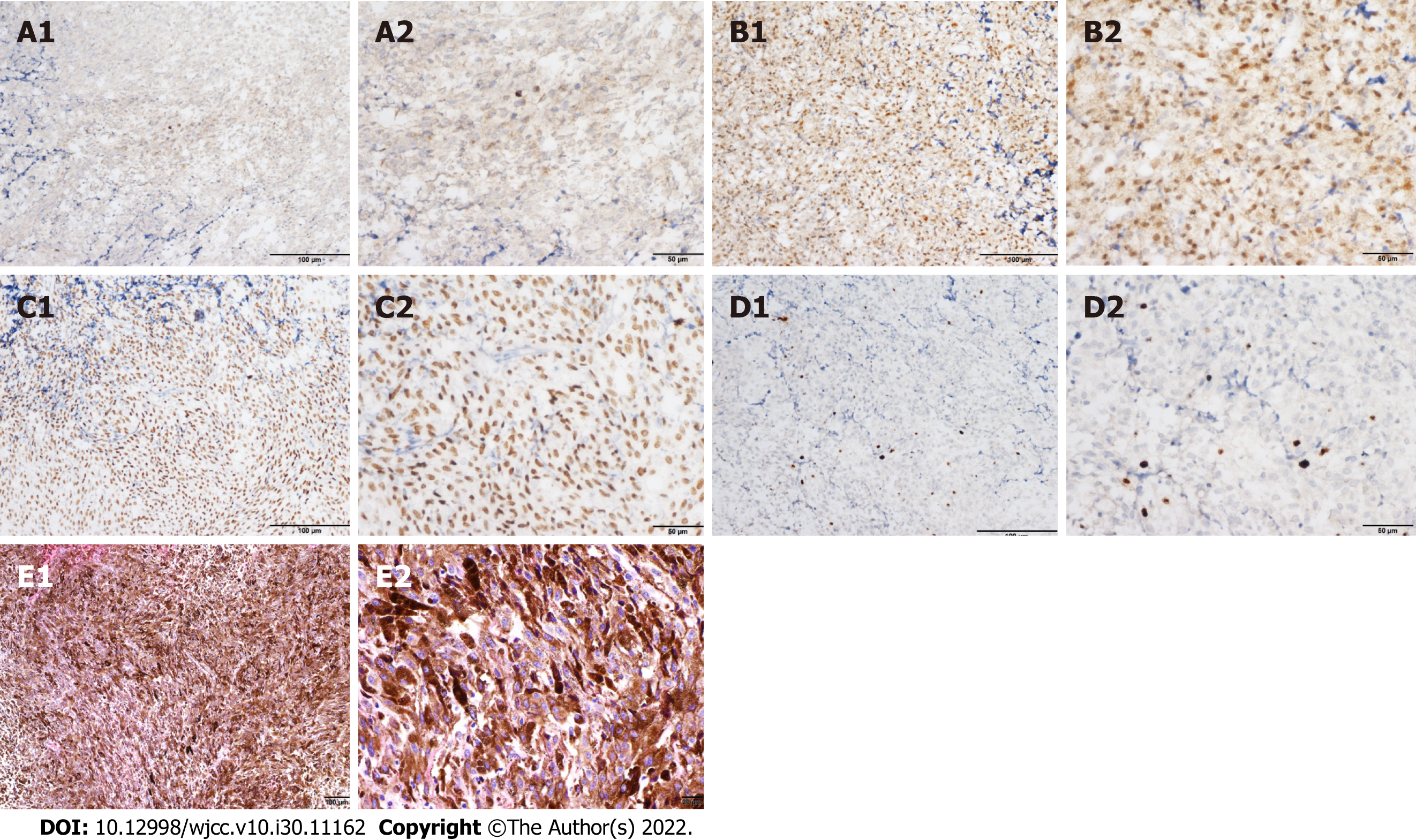
Figure 4 Immunohistochemical and histopathological analysis of confirmed recurrent melanoma at the 3rd operation.
A-D: Immunohistochemical staining of tumor cells showed positivity for (A) HMB-45, (B) S-100, (C) SOX-10, and (D) Ki67 (8%) (A1, B1, C1, and D1: × 200; A2, B2, C2, and D2: × 400); E: Hematoxylin-eosin staining showed that the tumor cells were diffusely distributed in flakes, with abundant cytoplasm, pigment granules in some cells, round or oval nuclei, nucleoli, and mitotic figures (3 cells/10 high power fields) (E1: × 100; E2: × 400).
- Citation: Wong TF, Chen YS, Zhang XH, Hu WM, Zhang XS, Lv YC, Huang DC, Deng ML, Chen ZP. Longest survival with primary intracranial malignant melanoma: A case report and literature review. World J Clin Cases 2022; 10(30): 11162-11171
- URL: https://www.wjgnet.com/2307-8960/full/v10/i30/11162.htm
- DOI: https://dx.doi.org/10.12998/wjcc.v10.i30.11162


