Copyright
©The Author(s) 2022.
World J Clin Cases. Oct 26, 2022; 10(30): 11155-11161
Published online Oct 26, 2022. doi: 10.12998/wjcc.v10.i30.11155
Published online Oct 26, 2022. doi: 10.12998/wjcc.v10.i30.11155
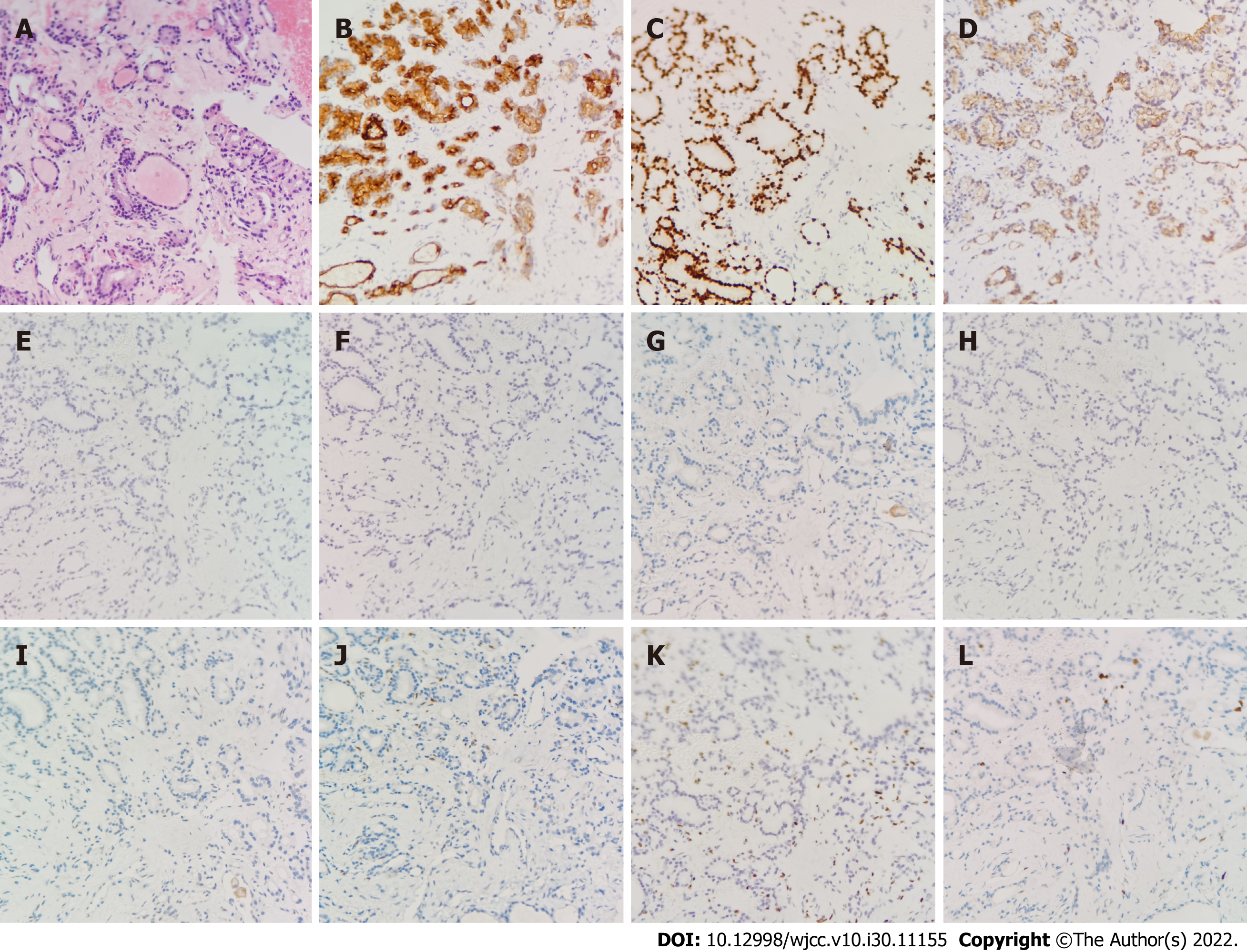
Figure 3 Hematoxylin-eosin staining and immunohistochemistry of percutaneous biopsy of the pelvic mass.
A: Hematoxylin-eosin staining showed multiple benign colloid-filled thyroid follicles (× 200); B-D: Immunohistochemistry (IHC) examination revealed that pelvic mass was positive for thyrobolulin (× 200), thyroid transcription factor-1 (× 200) and Cytokeratin-7 (× 200), respectively; E-L: IHC examination revealed that pelvic mass was negtive for Cytokeratin-20 (× 200), caudal-related homeobox transcription factor 2 (× 200), Estrogen receptor (× 200), calretinin (× 200), P53 (× 200), P16 (× 200), Wilms tumor-1 (× 200) and Ki-67 (× 200), respectively.
- Citation: Liu Y, Tang GY, Liu L, Sun HM, Zhu HY. Giant struma ovarii with pseudo-Meigs’syndrome and raised cancer antigen-125 levels: A case report. World J Clin Cases 2022; 10(30): 11155-11161
- URL: https://www.wjgnet.com/2307-8960/full/v10/i30/11155.htm
- DOI: https://dx.doi.org/10.12998/wjcc.v10.i30.11155


