Copyright
©The Author(s) 2022.
World J Clin Cases. Oct 26, 2022; 10(30): 11146-11154
Published online Oct 26, 2022. doi: 10.12998/wjcc.v10.i30.11146
Published online Oct 26, 2022. doi: 10.12998/wjcc.v10.i30.11146
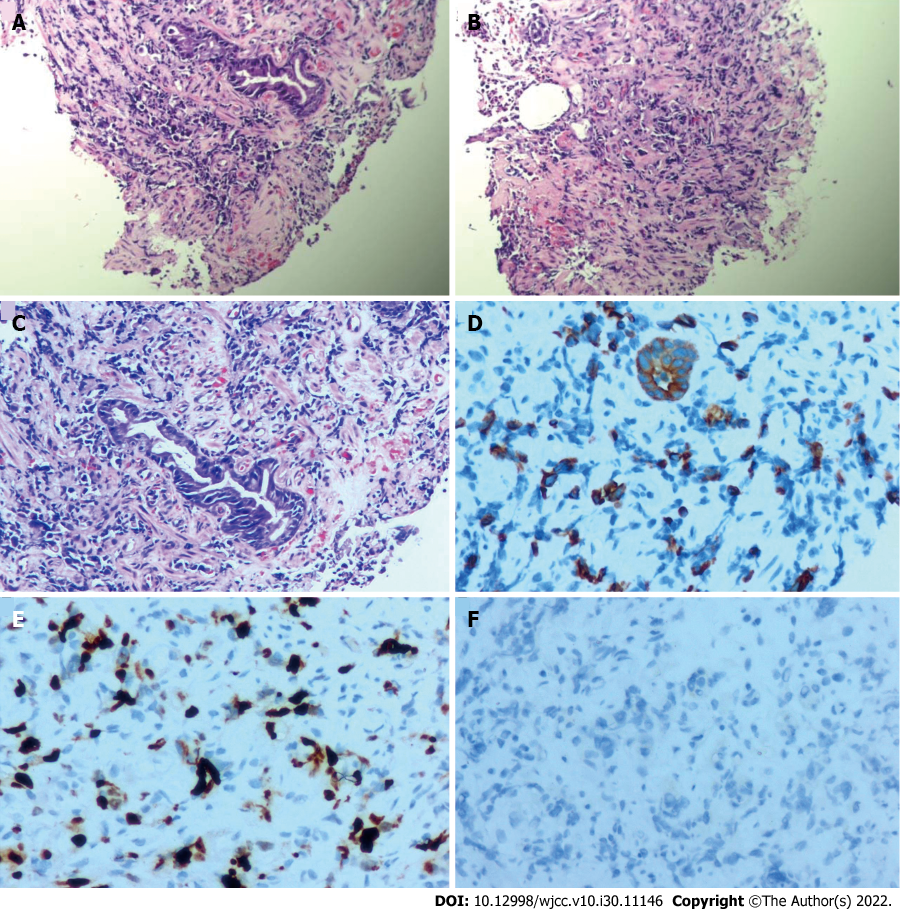
Figure 5 Pathological examination after gastroscopic biopsy reveals the presence of a small number of atypical cells.
A-C: Hematoxylin-eosin staining of biopsy specimens indicate poorly differentiated adenocarcinoma (A, 40 ×; B, 40 ×; C, 100 ×); D-F: Immunohistochemical staining shows that the specimens are positive for CKL (D, 200 ×), and Ki-65 (E, 200 ×) and negative for Her-2 (F, 200 ×).
- Citation: Zhao YX, Yang Z, Ma LB, Dang JY, Wang HY. Synchronous gastric cancer complicated with chronic myeloid leukemia (multiple primary cancers): A case report. World J Clin Cases 2022; 10(30): 11146-11154
- URL: https://www.wjgnet.com/2307-8960/full/v10/i30/11146.htm
- DOI: https://dx.doi.org/10.12998/wjcc.v10.i30.11146


